6 ANATOMY OF THE FEMALE PELVIS AND FETAL SKULL
CLINICALLY RELEVANT ANATOMY 123 ULNAR NERVE ENTRAPMENT2 PRECLINIC ACTIVITY – VOICE REVISION ANATOMY OF LARYNX
21 VISION BASIC ANATOMY AND PHYSIOLOGY OF THE VISUAL
23 NOISE EXPOSURE BASIC ANATOMY AND PHYSIOLOGY OF THE
4 THE ANATOMY OF SUCCESSFUL FEEDBACK CONFERENCING PRESENTER MARJORI
424010DOC ANATOMY AND PHYSIOLOGY OF THE EYE CONTRAST CONTRAST
6. Anatomy of the Female Pelvis and Fetal Skull
6. Anatomy of the Female Pelvis and Fetal Skull
Study Session 6 Anatomy of the Female Pelvis and Fetal Skull 3
Learning Outcomes for Study Session 6 3
6.1.3 Pubic bones and the symphysis pubis 5
6.2.1 The size and shape of the pelvis 6
6.3.4 Regions and landmarks in the fetal skull 12
Self-Assessment Questions (SAQs) for Study Session 6 13
SAQ 6.1 (tests Learning Outcomes 6.1, 6.2 and 6.3) 13
SAQ 6.2 (tests Learning Outcomes 6.1, 6.2 and 6.3) 15
Study Session 6 Anatomy of the Female Pelvis and Fetal Skull
Introduction
In this study session you will learn about the bony structures with the most importance for the pregnant woman and the baby she will give birth to. The bones of the skeleton have the main function of supporting our body weight and acting as attachment points for our muscles. The focus in this study session will be on the female pelvis, which supports the major load of the pregnant uterus, and the fetal skull, which has to pass through the woman’s pelvis when she gives birth.
There are certain key landmarks in the anatomy of the female pelvis and the fetal skull that we will show you in this study session. Knowing these landmarks will enable you to estimate the progress of labour, by identifying changes in their relative positions as the baby passes down the birth canal. You will learn how to do this in the next Module in this curriculum, which is on Labour and Delivery Care.
Learning Outcomes for Study Session 6
When you have studied this session, you should be able to:
6.1 Define and use correctly all of the
key words printed in bold.
(SAQs
6.1, 6.2 and 6.3)
6.2 Describe the female pelvis and identify the important features for obstetric care. (SAQs 6.1, 6.2 and 6.3)
6.3 Describe the main features of the fetal skull, and their importance for labour and delivery. (SAQs 6.1, 6.2 and 6.3)
6.1 The female bony pelvis
The pelvis is a hard ring of bone (see Figure 6.1), which supports and protects the pelvic organs and the contents of the abdominal cavity. The muscles of the legs, back and abdomen are attached to the pelvis, and their strength and power keep the body upright and enable it to bend and twist at the waist, and to walk and run.
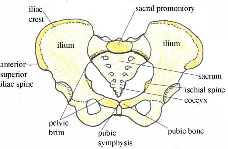
Figure 6.1 The bones of the female pelvis.
The woman’s pelvis is adapted for child bearing, and is a wider and flatter shape than the male pelvis. The pelvis is composed of pairs of bones, which are fused together so tightly that the joints are difficult to see. We will describe each of the bones in turn, and their major landmarks. It will help you to visualise the anatomy of the pelvis if you keep referring back to Figure 6.1.
6.1.1 Ilium
Ilium is pronounced ‘ill ee umm’ and iliac is ‘ill ee ack’.
The major portion of the pelvis is composed of two bones, each called the ilium — one on either side of the backbone (or spinal column) and curving towards the front of the body. When you place your hand on either hip, your hand rests on the iliac crest, which is the upper border of the ilium on that side. At the front of the iliac crest, you can feel the bony protuberance called the anterior superior iliac spine (a ‘protuberance’ is something that sticks out, like a little hill or knob).
Question
What do the directional terms ‘anterior superior’ tell you about the position of the iliac spines? (If you can’t remember, look back at Box 3.1 in Study Session 3.)
Answer
Anterior tells you that the iliac spines are at the front of the body, and superior tells you that they are above the main portion of the ilium on each side.
End of answer
6.1.2 Ischium
The ischium is the thick lower part of the pelvis, formed from two fused bones — one on either side. When a woman is in labour, the descent of the fetal head as it moves down the birth canal is estimated in relation to the ischial spines, which are inward projections of the ischium on each side. The ischial spines are smaller and rounder in shape in the woman’s pelvis than in that of the man. In the Module on Labour and Delivery Care, you will learn how to feel for the ischial spines to help you estimate how far down the birth canal the baby’s head has progressed.
Ischium is pronounced ‘iss kee umm’ and ischial is ‘iss kee al’.
6.1.3 Pubic bones and the symphysis pubis
The pubic bones on either side form the front part of the pelvis. The two pubic bones meet in the middle at the pubic symphysis. (A symphysis is a very strong bony joint.) The pubic symphysis is immediately below the hair-covered pubic mound that protects the woman’s external genitalia (as shown in Figure 3.2, if you want to look back at it now).
When you examine the abdomen of a pregnant woman, feeling for the top of the pubic symphysis with your fingers is a very important landmark. In Study Session 10, you will learn how to measure the height of the uterus from the pubic symphysis to the fundus (top of the uterus — see Figure 3.3 if you need to remind yourself of the position of the fundus). This measurement enables you to estimate the gestational age of the fetus, i.e. how many weeks of the pregnancy have passed, and whether the fetus is growing at the normal rate.
6.1.4 Sacrum
Sacrum is pronounced ‘say krum’. Coccyx is pronounced ‘kok six’.
The sacrum is a tapered, wedge-shaped bone at the back of the pelvis, consisting of five fused vertebrae (the small bones that make up the spinal column or backbone). At the bottom of the sacrum is a tail-like bony projection called the coccyx. The upper border of the first vertebra in the sacrum sticks out, and points towards the front of the body; this protuberance is the sacral promontory — an important landmark for labour and delivery.
6.2 The pelvic canal
The roughly circular space enclosed by the pubic bones at the front, and the ischium on either side at the back, is called the pelvic canal — the bony passage through which the baby must pass. This canal has a curved shape because of the difference in size between the anterior (front) and posterior (back) borders of the space created by the pelvic bones. You can see it from the side view in Figure 6.2.
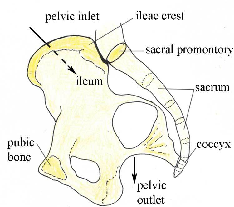
Figure 6.2 The pelvic canal seen from the side, with the body facing to the left.
6.2.1 The size and shape of the pelvis
The size and shape of the pelvis is important for labour and delivery. Well-built healthy women, who had a good diet during their childhood growth period, usually have a broad pelvis that is well adapted for childbirth. It has a round pelvic brim and short, blunt ischial spines. (Doctors and midwives call this shape a ‘gynaecoid’ pelvis.) It gives the least difficulty during childbirth, provided the fetus is a normal size and the birth canal has no abnormal tissue growth causing an obstruction.
There is considerable variation in pelvis shapes, some of which create problems in labour and delivery. A narrow pelvis can make it difficult for the baby to pass through the pelvic canal. A deficiency of important minerals like iodine in the diet during childhood may result in abnormal development of the pelvic bones. Stunting (being much shorter than average for age) due to malnutrition and/or infectious diseases can also result in a narrow pelvis.
Next, we look at the shape of the pelvic canal in more detail, and distinguish between the pelvic inlet (the roughly circular space where the baby’s head enters the pelvis — Figure 6.3), and the pelvic outlet (the roughly circular space where the baby’s head emerges from the pelvis. As you will see in the next section, the inlet and the outlet of the pelvis are not the same size.
6.2.2 The pelvic inlet
The pelvic inlet is formed by the pelvic brim, which you saw in Figure 6.1. The pelvic brim is rounded, except where the sacral promontory and the ischial spines project into it. The dimensions in centimetres (cm) of the pelvic inlet are shown in Figure 6.3 in both directions (top to bottom; and transverse or side to side). When you look at Figure 6.3, imagine that you are a baby in the head-down position, looking down on the pelvis from above, at the space you must squeeze through! It is just 13 cm wide (on average) and 12 cm from top to bottom.
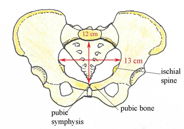
Figure 6.3 Diameters of the pelvic inlet, viewed from above.
6.2.3 The pelvic outlet
The pelvic outlet is formed by the lower border of the pubic bones at the front, and the lower border of the sacrum at the back. The ischial spines point into this space on both sides. Figure 6.4 shows the dimensions of the space that the fetus must pass through as it emerges from the mother’s pelvis. As you look at Figure 6.4, imagine that you are the birth attendant who is looking up the birth canal, waiting to see the fetal head emerging.
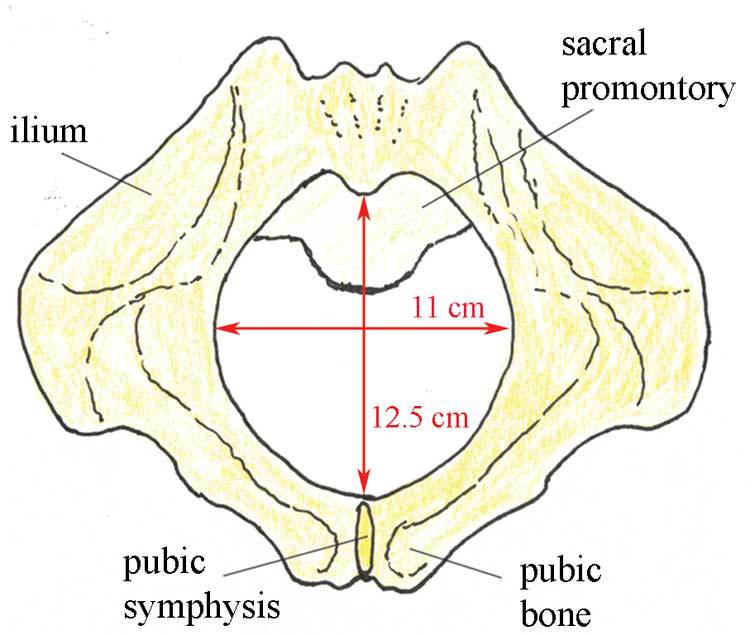
Figure 6.4 Diameters of the pelvic outlet, viewed from below.
Question
What do you notice when you compare the dimensions of the pelvic inlet (Figure 6.3) and the pelvic outlet (Figure 6.4)? Which is the narrowest?
Answer
The narrowest diameter for the fetus to pass through is the pelvic outlet, which is only 11 cm wide in the average female pelvis.
End of answer
It is difficult to see from Figures 6.3 and 6.4, but the fetus has to rotate in order to get through the pelvic canal. This is because the pelvic inlet is 13 cm wide, whereas the pelvic outlet is only 11 cm wide. In order to fit through the pelvic outlet at its widest dimension (12.5 cm from top to bottom), the fetus must rotate so it ‘presents’ its head to the widest dimension of the pelvic cavity at every point as it passes through. The largest part of the fetus is the skull, so the baby’s head rotates first, and the shoulders and the rest of the body follow. You will learn all about this in the Labour and Delivery Care Module. First, we have to look more closely at the structure of the fetal skull.
6.3 The fetal skull
The fetal skull is the most difficult part of the baby to pass through the mother’s pelvic canal, due to the hard bony nature of the skull. Understanding the anatomy of the fetal skull and its diameter will help you recognise how a labour is progressing, and whether the baby’s head is ‘presenting’ correctly as it comes down the birth canal. This will give you a better understanding of whether a normal vaginal delivery is likely, or if the mother needs referral because the descent of the baby’s head is not making sufficient progress.
6.3.1 Fetal skull bones
The skull bones encase and protect the brain, which is very delicate and subjected to pressure when the fetal head passes down the birth canal. Correct presentation of the smallest diameter of the fetal skull to the largest diameter of the mother’s bony pelvis is essential if delivery is to proceed normally. But if the presenting diameter of the fetal skull is larger than the maternal pelvic diameter, it needs very close attention for the baby to go through a normal vaginal delivery.
You can locate the main skull bones in Figure 6.5.
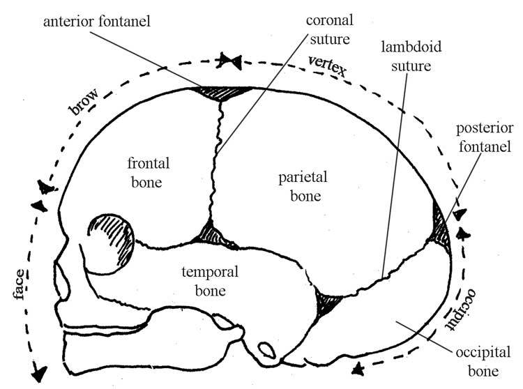
Figure 6.5 Bones of the fetal skull — side view facing left.
The fetal skull bones are as follows:
The frontal bone, which forms the forehead. In the fetus, the frontal bone is in two halves, which fuse (join) into a single bone after the age of eight years.
The two parietal bones, which lie on either side of the skull and occupy most of the skull.
Parietal is pronounced ‘parr eye ett al’. Occipital is pronounced ‘ox ipp itt al’.
The occipital bone, which forms the back of the skull and part of its base. It joins with the cervical vertebrae (neck bones in the spinal column, or backbone).
The two temporal bones, one on each side of the head, closest to the ear.
Understanding the landmarks and measurements of the fetal skull will help you to recognise normal and abnormal presentations of the fetus during antenatal examinations, labour and delivery.
6.3.2 Sutures
Sutures are joints between the bones of the skull. In the fetus they can ‘give’ a little under the pressure on the baby’s head as it passes down the birth canal. During early childhood, these sutures harden and the skull bones can no longer move relative to one another, as they can to a small extent in the fetus and newborn. It is traditional for their names and locations to be taught in midwifery courses. You may be able to tell the angle of the baby’s head as it ‘presents’ in the birth canal by feeling for the position of the main sutures with your examining fingers. You can see the position of the sutures in the fetal skull in Figure 6.6, and also the diameters at two points.
Suture is pronounced ‘soo tyor’.
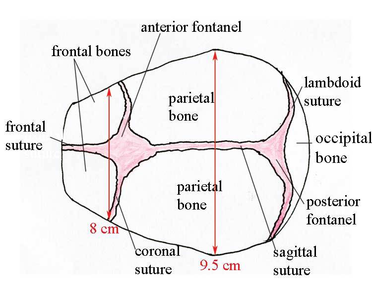
Figure 6.6 Regions and landmarks in the fetal skull facing to the left, as seen from above. Notice the average diameters in red.
The lambdoid suture forms the junction between the occipital and the frontal bone.
Lambdoid is pronounced ‘lamm doyd’. Sagittal is ‘saj itt al’ and coronal is ‘korr oh nal’.
The sagittal suture joins the two parietal bones together.
The coronal suture joins the frontal bone to the two parietal bones.
The frontal suture joins the two frontal bones together.
Question
What do you notice about the diameters given in Figure 6.6, relative to the dimensions of the pelvic canal (Figures 6.3 and 6.4)?
Answer
At its widest part, the fetal skull is (on average) 9.5 cm wide. This is 3.5 cm less than the widest diameter of the pelvic inlet, and 1.5 cm less than the widest diameter of the pelvic outlet.
End of answer
Thus, if the mother’s pelvis and the fetal skull are the average size, there is just sufficient room for the baby’s head to pass through the pelvic canal if the head rotates to present to the widest dimension of the pelvis.
6.3.3 Fontanels
A fontanel is the space created by the joining of two or more sutures. It is covered by thick membranes and the skin on the baby’s head, protecting the brain underneath the fontanel from contact with the outside world. Identification of the two large fontanels on the top of the fetal skull helps you to locate the angle at which the baby’s head is presenting during labour and delivery. The fontanels are shown in Figures 6.5 and 6.6. They are:
The anterior fontanel (also known as the bregma) is a diamond-shaped space towards the front of the baby’s head, at the junction of the sagittal, coronal and frontal sutures. It is very soft and you can feel the fetal heart beat by placing your fingers gently on the fontanel. The skin over the fontanel can be seen ‘pulsing’ in a newborn or young baby.
The posterior fontanel (or lambda) has a triangular shape, and is found towards the back of the fetal skull. It is formed by the junction of the lambdoid and sagittal sutures.
6.3.4 Regions and landmarks in the fetal skull
Figures 6.5 and 6.6 allow you to identify certain regions and landmarks in the fetal skull, which have particular importance for obstetric care because they may form the so-called presenting part of the fetus — that is, the part leading the way down the birth canal.
The vertex is the area midway between the anterior fontanel, the two parietal bones and the posterior fontanel. A vertex presentation occurs when this part of the fetal skull is leading the way. This is the normal and the safest presentation for a vaginal delivery.
The brow is the area of skull which extends from the anterior fontanel to the upper border of the eye. A brow presentation is a significant risk for the mother and the baby.
The face extends from the upper ridge of the eye to the nose and chin (lower jaw). A face presentation is also a significant risk for the mother and baby.
The occiput is the area between the base of the skull and the posterior fontanel. It is unusual and very risky for the occiput to be the presenting part.
When you study the next Module on Labour and Delivery Care, you will learn about other presentations, including ‘breech’ (the baby is head-up and its feet or bottom is the presenting part), and ‘shoulder’ first.
Now that you know all the major anatomical features of the female reproductive system, the female pelvis and the fetal skull, we move on in Study Session 7 to consider the major physiological changes that take place in a woman’s body during pregnancy.
Summary of Study Session 6
In Study Session 6, you have learned that:
The bony pelvis is composed of the ilium, ischium, pubic bones and sacrum.
The size and shape of the bony pelvis can affect the ease or difficulty of labour and delivery; a broad pelvis gives less difficulty than a narrow one, which may obstruct the descent of the baby down the birth canal.
Certain landmarks in the anatomy of the pelvis are commonly used to estimate the descent of the baby during labour and delivery. The two most important landmarks are the ischial spines and the sacral promontory, which can be felt with the fingers during a vaginal examination.
The pelvic inlet is the space where the baby’s head enters the pelvis; it is larger than the pelvic outlet, where the baby’s head emerges from the pelvis. In order to get through the widest diameter of the inlet and the outlet, the baby has to rotate as it passes through the pelvic canal.
The skull is formed by several bones joined tightly together by joints called sutures. In the fetus and newborn, spaces called fontanels exist between some of the skull bones on the top of the baby’s head. The position of the sutures and the fontanels can tell you about the angle at which the baby’s head is presenting during labour and delivery.
The vertex presentation (where the top of the baby’s head is the presenting part) is the most common and the safest presentation for a normal vaginal delivery. Other presentations carry a much higher risk for the mother and baby.
Self-Assessment Questions (SAQs) for Study Session 6
Now that you have completed this study session, you can assess how well you have achieved its Learning Outcomes by answering these questions. Write your answers in your Study Diary and discuss them with your Tutor at the next Study Support Meeting. You can check your answers with the Notes on the Self-Assessment Questions at the end of this Module.
SAQ 6.1 (tests Learning Outcomes 6.1, 6.2 and 6.3)
Match each anatomical name with the correct description.
Matching quiz
Options
Fused vertebrae at the back of the bony pelivs
![]()
Hip bone in the pelvis
![]()
Joint between the parietal bones in the fetal skull
![]()
Paired bones forming the front of the skull
![]()
The top of the fetal skull between the two fontanels
![]()
Matches
Frontal bones
![]()
Ilium
![]()
Sacrum
![]()
Sagittal suture
![]()
Vertex
![]()
Solution
Pair 1
Hip bone in the pelvis
![]()
Ilium
![]()
Pair 2
Paired bones forming the front of the skull
![]()
Frontal bones
![]()
Pair 3
Joint between the parietal bones in the fetal skull
![]()
Sagittal suture
![]()
Pair 4
Fused vertebrae at the back of the bony pelivs
![]()
Sacrum
![]()
Pair 5
The top of the fetal skull between the two fontanels
![]()
Vertex
![]()
SAQ 6.2 (tests Learning Outcomes 6.1, 6.2 and 6.3)
Which of the following statements is false? In each case, say why it is incorrect.
A The female bony pelvis is broader and flatter than the male pelvis.
B The pelvic inlet is narrower than the pelvic outlet.
C The iliac crest is an important landmark in measuring the progress of the fetus down the birth canal.
D The sutures in the fetal skull are strong hard joints that hold the skull bones rigidly in place.
E A newborn baby’s pulse can be seen beating in the anterior fontanel.
Answer
A is true. The female bony pelvis is broader and flatter than the male pelvis.
B is false. The pelvic inlet is wider (not narrower) than the pelvic outlet.
C is false. The iliac crest is the protuberance at the front of each hip bone; it is not important in measuring the progress of the fetus down the birth canal.
D is false. The sutures in the fetal skull ‘give’ a little under the pressure in the birth canal, allowing the skull bones to move to a small extent. This makes it easier for the baby’s head to pass through the mother’s bony pelvis.
E is true. A newborn baby’s pulse can be seen beating in the anterior fontanel.
End of answer
SAQ 6.3 (tests Learning Outcomes 6.1, 6.2 and 6.3)
List four possible features of the maternal bony pelvis and/or the fetal skull that may result in a difficult labour and delivery.
Answer
The possible features of the maternal bony pelvis and/or the fetal skull that may result in a difficult labour and delivery include (you only had to suggest four):
A narrow or deformed pelvis
Abnormal growth of tissue in the pelvic cavity
A large fetal skull
A brow, face, breech or shoulder presentation of the fetus
A fetus that does not present the widest part of its skull to the widest part of the pelvic inlet, and then rotate to do the same in the pelvic outlet.
End of answer
6 ANATOMY OF THE FEMALE PELVIS AND FETAL SKULL
ANATOMY & PHYSIOLOGY 166 SPRING 2010 COURSE INFORMATION OBJECTIVES
ANATOMY & PHYSIOLOGY EYES & EARS OUTLINE FOR NOTES
Tags: anatomy of, the anatomy, female, skull, fetal, anatomy, pelvis
- NAMES ES 342 CHAPTER 2 ELECTROMAGNETIC
- TOP OF FORM BEK NR 218 AF 30032005 GÆLDENDE
- APLICACIÓN INMEDIATA DE NORMAS FAVORABLES LA APLICACIÓN INMEDIATA DE
- 1 WWWASESORIAFUNDACIONESYASOCIACIONESES QUITAR EN WWWASESORIAFUNDACIONESYASOCIACIONESESINDEX AL FINAL DE LAS
- DCS EDUCATION SERVICES TRAININGS OFFERED IN AN EFFORT
- ¿LOS EQUIPOS DE TRABAJO VIRTUALES SON REALES? REFLEXIONES E
- 8 RDJ1382009 CONTRALORÍA GENERAL DE LA REPÚBLICA DIVISIÓN
- INMIGRACIÓN Y SERVICIOS SOCIALES ENTRE EL ESTEREOTIPO Y LA
- PROJECTBASED LEARNING EVALUATION INFORMATION FOR PARENTS THE CENTER FOR
- Le Futur Qu’estce que C’est? In French When we
- EK1 (DEĞIŞIKRG113200927166) ÖZEL HASTANELERDE ASGARÎ BULUNDURULACAK SAĞLIK PERSONELİ IMESUL
- Karta Zgłoszenia Międzynarodowa Konferencja Naukowa Uczenie się Przez Całe
- 8 FRAGEBOGEN FÜR PFLEGER ZUM THEMA LEBENSQUALITÄT BEI DER
- 8 PERATURAN KEPALA ARSIP NASIONAL REPUBLIK INDONESIA NOMOR 47
- A DVANCED NAVIGATION 28TH 29TH APRIL 2012 VAGABOND’S
- 2008 OUT OF STATE COLLEGE APPLICATION ESSAYS COMMON APPLICATION
- EQUAL OPPORTUNITY PROGRAM INSPECTION CHECKLIST EQUAL OPPORTUNITY PROGRAM INSPECTION
- PATVIRTINTA ŠVIETIMO KULTŪROS IR SPORTO SKYRIAUS VEDĖJO 2020 M
- NOTES FOR GUIDANCE TO BE USED ONLY BY TRAINED
- GUÍA DEL CANDIDATO – MUNDUS LINDO SEGUNDA COHORTE INTRODUCCIÓN
- CURRICULUM VITAE FRITZ RENDEMANN PERSONAL DATA ADDRESS AMAGERGADE
- 8 FELSŐOKTATÁSI INTÉZMÉNY NEVE KÖZOKTATÁSI INTÉZMÉNY NEVE ÉS OM
- SEXUAL ABUSE AGAINST CHILDREN IN INSTITUTIONS – STATISTICS STUDIES
- QOSTIC39 R1 ASSOCIATION FRANÇAISE DES UTILISATEURS DE TÉLÉCOMMUNICATIONS
- 8 PATVIRTINTA ŠILUTĖS RAJONO SAVIVALDYBĖS TARYBOS 2021 M D
- OPIS MODUŁU KSZTAŁCENIA ZAŁĄCZNIK NR NAZWA MODUŁU (PRZEDMIOTU) PRAKTYKA
- BASE DE DATOS CONVOCATORIAS ABIERTAS COOPERACION INTERNACIONAL SEPTIEMBRE DE
- TEMARIO REVISADO DE LENGUA CASTELLANA Y LITERATURA 1 LA
- APPENDIX 6 FILE METERSDOC MULTIMETERS A MULTIMETERS IS THE
- C O L E G I O P R
ESSENTIAL STUDENT UNDERSTANDINGS KINDERGARTEN THIS EXAMPLE IS FROM ANOTHER
ARRAIGADOS Y EDIFICADOS EN CRISTO FIRMES EN LA FE”
CAIET DE SARCINI OFERTA CAZARE PACHET CAZARE CĂMIN VIII
WTGCW572 PÁGINA 0 ORGANIZACIÓN MUNDIAL DEL COMERCIO WTGCW581 29
BURSA DE VALORI BUCURESTI SA REINFIINTAT LA DATA DE
PREFERRED LENDER PROGRAM CREDIT MANAGEMENT SYSTEM SUMMARY THE FOLLOWING
 CONVENIOS 2015 RESERVAS SUSANA GONZALEZ EJECUTIVA DE VENTAS TELÉFONO
 PRÉFECTURE DE LA RÉUNION ARRETE N° 1952DRASS PORTANT MODIFICATION
YEAR COMPANY PROJECT NAME 20056 ADVANCE PARTY FILMS RED
ODVOZ KUĆNOG OTPADA U VRIJEME BLAGDANA OBAVJEŠTAVAMO STANOVNIKE
14 FEATURE ECONOMY IN SOUND SYSTEMS NICK CLEMENTS LABORATOIRE
REVISED JAN 2012 KEARNEY AGRICULTURAL RESEARCH AND EXTENSION CENTER
 FO 2502 FALLANALYSE 23 PHASE VA ANLEITUNG FALLANALYSE
 ÍNDICE LA ENCUESTA 3 CONCEPTO 4 TIPOS DE ENCUESTAS
6 SZENTMÁRTONKÁTA ÖNKORMÁNYZAT KÉPVISELŐTESTÜLETE H E L Y B
BRAIN DEATH AND CARE OF THE ORGAN DONATION PATIENT
PODACI O RAJONIMA ZA OBAVLJANJE DIMNJAČARSKIH POSLOVA NA PODRUČJU
 PROCEEDINGS OF THE 61ST INTERNATIONAL CONVENTION OF SOCIETY OF
 EXPLORANDO MATERIALES GRAFENO ¡INTENTA ESTO! 1 TOMA UN
ETIKA 1 HOMO FABER MOĆ TIŠINA AKO BOG NE
