ANPS 019 BENEYTOSANTONJA 113012 SPECIAL SENSES II AUDITORY
ANPS 019 BENEYTOSANTONJA 113012 SPECIAL SENSES II AUDITORY
ANPS 019 Beneyto-Santonja 11/30/12
Special Senses II Auditory & Vestibular Systems
What is the anatomy of the ear?
External ear – sound collection
Middle ear – sound amplification
Inner ear – sound detection (cochlea) & balance (vestibular apparatus)
External Ear
Auricle: provides directional sensitivity
Tympanic Membrane (eardrum): separates external ear from middle ear
Middle Ear aka Tympanic Cavity
Auditory Ossicles – 3 smallest bones in the body:
Malleus (hammer), Incus (anvil), Stapes (stirrup)
Malleus attached to tympanic membrane
Stapes attached to oval window of cochlea
Two smallest muscles in body protect ear from prolonged loud sounds:
Tensor Tympani – stiffens tympanic membrane
Stapedius – reduces movement of stapes at oval window
Eustachian tube: equalizes pressure within middle ear
Inner Ear
Subdivided into:
Vestibule – balance
Semicircular canals – balance
Cochlea – auditory
Vibration of Tympanic Membrane
Converts sound waves at tympanic membrane into movement of fluids in membranous labyrinth of cochlea
Auditory receptors lie within the Organ of Corti of the cochlea
Organ of Corti
Hair cells = mechanoreceptors
The Organ of Corti rests on the basilar membrane
The auditory receptors, known as Hair cells, have cilia that are in contact with the tectorial membrane
Movement of the basilar membrane causes movement of the cilia and depolarization of the hair cells
How does the brain perceive sound?
Sound frequency is mapped on the basilar membrane
The map is maintained in the cochlear nerve and cochlear nucleus in the brainstem
The map is maintained all the way to the auditory cortex in the temporal lobe
Auditory Pathway
Synapse in Cochlear nuclei in medulla
Synapse in inferior colliculus
Synapse in medial geniculate nucleus of thalamus
Synapse in auditory cortex of temporal lobe
How does the ear help with balance?
Vestibular System
The vestibular apparatus in the inner ear consists of two Otolith Organs:
Utricle: responds to tilting and horizontal movement (moving car)
Saccule: responds to vertical movement (elevator)
Semicircular Canals detect rotation
There is one semicircular canal for each plane of space
Gustation and Olfaction (Taste and Smell)
How are smell and taste clinically important?
Taste intimately linked to sense of smell
The number of taste buds begins declining rapidly by age 50
Sense of smell declines with ageing
Elderly aren’t motivated to eat because food has little taste
Parkinson’s Anosmia
In Parkinson’s disease, the incidence of smell loss is greater than the incidence of tremor.
Taste buds
Gustatory Receptors = Chemoreceptors
Clustered in papillae of tongue
Gustatory receptors are specialized epithelial cells NOT neurons
Survive only 10 days before replacement
Taste Zones do not correspond to papillae types
Primary: sweet, salty, sour, bitter
Additional
Umami
Receptors sensitive primarily to amino acids
Characteristic of beef/chicken broths and parmesan cheese
Water
Detected by receptors in the pharynx (‘dry throat’)
Fats
Olfactory Receptors = Chemoreceptors
Olfactory epithelium contains:
Olfactory receptors = bipolar neurons
Basal (stem) cells
One of few examples of neurons replaced throughout life
Olfactory Pathway
Olfactory tract has 3 primary targets:
Contralateral olfactory bulb – smell localization
Thalamus – smell perception
Limbic system WITHOUT A THALAMIC RELAY – smell emotion
The Olfactory system project to the Limbic system without a thalamic relay
Tags: special, beneytosantonja, auditory, 113012, senses
- GUÍA DE AUTOEVALUACIÓN PARA CARRERAS DE ARQUITECTURA SISTEMA ARCUSUR
- ACUERDO DE ENCOMIENDA DE GESTIÓN ENTRE EL MINISTERIO DE
- INFORME RAONAT SOBRE LA IDONEÏTAT DEL MEMBRE PROPOSAT PER
- ZAŁĄCZNIK DO UCHWAŁY NR KEXVI00071512016 RADY GMINY DĘBE WIELKIE
- KAFKAS ÜNİVERSİTESİ ATATÜRK SAĞLIK HİZMETLERİ MESLEK YÜKSEKOKULU STRATEJİK PLAN
- 4 SECTION VIII PLAN GENERAL PROVISIONS
- İHKİB KAĞITHANE MESLEKİ VE TEKNİK ANADOLU LİSESİ TEKSİL TEKNOLOJİ
- NOTES FOR NEWSLETTER WINTER 1997 VOL 6 NO 1
- LA TORRECILLA S ITUACIÓN EL CASTILLO DEL ALCORLO TAMBIÉN
- ACUERDO NÚMERO DIECISIETE GUIÓN DOS MIL CATORCE (172014) GUATEMALA
- KEK KH TÁJÉKOZTATÓ AZ ELEKTRONIKUS ÜGYINTÉZÉSRŐL ELEKTRONIKUS NYOMTATVÁNY KITÖLTÉSÉRŐL
- MEGHÍVÓ TISZTELT ÁLLÁSKERESŐ! SZERETETTEL VÁRJUKA VÁLLALKOZZ ITTHON FIATAL! PROGRAM
- CONTRACT FOR CONDUCT OF A GRAND PRIX REGATTA WE
- UFFICIO NOTARILE ASSOCIATO – ESECUZIONI IMMOBILIARI TRIBUNALE DI PARMA
- PROGRAMA “LOS NOCTURNOS” INTRODUCCIÓN FEBE (FEDERACIÓN ESPAÑOLA DE BEBIDAS
- ACUERDO ENTRE LA ADMINISTRACIÓN DE LA COMUNIDAD FORAL DE
- C ONSULATE GENERAL OF THE CZECH REPUBLIC & IN
- ACTIVIDAD GRUPAL 1) PRESENTACIÓN DE LOS COORDINADORES (PADRES
- “SUPUESTOS ADICIONALES Y PRÁCTICAS DE CONTABILIDAD FINANCIERA” PROFESORA MARÍA
- SIGNIFICANT CHANGES TO THE GRANT GENERAL CONDITIONS (GC1) DATED
- ACUERDO SOCIAL PARA LA PRODUCTIVIDAD Y EL EMPLEO EN
- Nº 84 EN LA CIUDAD DE RESISTENCIA CAPITAL
- SZKOLNY PROGRAM WSPIERANIA UCZNIA ZE SPECJALNYMI POTRZEBAMI EDUKACYJNYMI SZKOŁA
- ACTIVIDADES DE REFUERZO DE MATEMÁTICAS 1º ESO TEMA 2
- D2185 YİM 8084 YÜKSEK İDARE MAHKEMESI OLARAK OTURUM YAPAN
- ACUERDO DE LAS CONDICIONES DE TRABAJO DE LOS EMPLEADOS PÚBLICOS DE LA ADMINISTRACIÓN LOCAL DE
- TRANSMITTED BY THE EXPERT FROM OICA INFORMAL DOCUMENT NO
- ACTION PLAN TEMPLATE FOR MAINTAINING ESSENTIAL SERVICEACTIVITY KEY CUSTOMERS
- UNIVERSIDADE METODISTA DE ANGOLA LISTAGEM DE REVISTAS E JORNAIS
- EJERCICIOS I ESCRIBE CON LAS CONECTIVAS Y SÍMBOLOS DE
CO WARTO WIEDZIEĆ O ZNIECZULENIU ? I INFORMACJE OGÓLNE
 USULAN DAN MUTASI PESERTA PBI APBD KOTA BOGOR SOP
THOMAS OWEN CÉRÉMONIAL NOCTURNE (1966) MON PÈRE NE M’IMPOSAIT
SAINT PAUL PUBLIC SCHOOLS HUMAN RESOURCES WORKFORCE TEAM THE
15A NCAC 18C 0405 STORAGE OF FINISHED WATER (A)
TEXAS FORUM FOR REPRODUCTIVE SCIENCES 26TH ANNUAL VIRTUAL MEETING
UVOD U BAZE PODATAKA BAZE PODATAKA PREDSTAVLJAJU VIŠU RAZINU
 CHAPTER 3 INTERPOLATION AND POLYNOMIAL APPROXIMATION 31 INTERPOLATION
 UNIT HEALTH AND SAFETY STRUCTURE HEAD OF THE
Ðïࡱáþÿ ¦âþÿÿÿ¤¥¥áo Ø¿x88tbjbj¸ý¸ý x9f²úx97zfúx97zf Ÿÿÿÿÿÿ·^^ôôôôôÿÿÿÿ8x9cüx84¨l`â9)¤ý)d!ßááááááô¶ªcx8c]ôe)9)eeôôõb¡¡¡e^ôôß¡eß¡¡ªù9|åÿÿÿÿ0(ëx9dxõÿÿÿÿ£uëx0¨g~6dñ6d
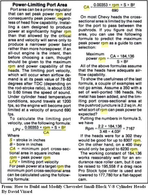 THIS CALCULATION IS USED TO ESTIMATE THE MAXIMUM RPM
1629 EN MATERIA DE PRESCRIPCIÓN LAS ACCIONES PENALES POR
 1 CASO ESTUDIO ENSAYO CLÍNICO RANDOMISED TRIAL OF CHOLESTEROL
INICIATIVAS PRESENTADAS EN LAS CORTES DE ARAGÓN SOBRE LA
 REGLAMENTO DE LA LEY DEL IMPUESTO ESPECIAL SOBRE PRODUCCIÓN
APELLIDOS Y NOMBRE SANZ GARCÍA Mª AGRIPINA Nº
CONFIDENCIALIDAD DERECHO A ELEGIR Y CONSENTIMIENTO LA CONFIDENCIALIDAD
 AÑO 40 ENERO 1998 NO 837 ND SACERDOTE PAULISTA
LỊCH LÀM VIỆC TRUNG TÂM KIỂM NGHIỆM DP MP
 SPONSORS MICROCHIP TECHNOLOGY IS THE ORGANIZING SPONSOR FOR THE
