NEW YORK SCIENCE JOURNAL 2010 3(11) ASPECTS OF THE
BIOLOGY COMBINED SCIENCE GCSE (9–1) GATEWAYBIS SCIENCE AND SOCIETY COMMUNITY CHALLENGE GRANT SCHEME
CHARLES UNIVERSITY OF PRAGUE FACULTY OF SCIENCE
DEPARTMENT OF CROP AND SOIL SCIENCE RECRUITMENT INFORMATION
DEPARTMENT OF EDUCATION AND SCIENCE SUMMARY OF ALL
FACULTY OF ENGINEERING AND PHYSICAL SCIENCES TAUGHT
Aspects of reproductive biology in mudskipper, Periophthalmus papilio from mangrove swamps of Lagos lagoon, Lagos, Nigeria
New York Science Journal 2010; 3(11)
Aspects of the reproductive biology in mudskipper, Periophthalmus papilio from mangrove swamps of Lagos lagoon, Lagos, Nigeria.
LAWSON Emmanuel O.
Department of Fisheries, Faculty of Science, Lagos State University, Ojo
P.O. Box 001, LASU Post Office, Lagos, Nigeria.
Abstract: The present study was conducted on aspects of reproductive biology of mudskipper, Periophthalmus papilio (Bloch and Schneider 1801) from the mangrove swamps of Lagos lagoon in Nigeria. P. papilio is an economically valued fish species, both as food for the populace and baits for industrial or offshore fisheries in Nigeria and the entire coasts of West African sub-region. 2167 individual fish were caught from the lagoon with non return valve traps between May 2006 and May 2008. Data on sex, total length (TL), gonad (GW) body weight (BW) measurements were carried out on individual specimen. These data were used in the determination of sex ratio, gonadosomatic index (GSI), and maturation in this species. The sex ratio of 1 male to 1.42 females was observed but was not significantly different (X2=57.04>X21, 0.05=3.84) but a departure from the theoretical 1male:1female sex ratio. Females population were more than the males. The GSI values varied from 0.01 to 0.48% in males and between 0.11 and 8.40% in females. 0.104±0.004 (in males) and 2.69±0.03% (in females) of the body mass were used in gonad development. The eggs were as small as 0.20mm and as big as 0.50 mm (mean = 0.36±0.01 mm) in diameter for average fish of 116 mm TL.. Seven (7) stages of maturity: immature, immature and developing, ripening, ripe, ripe running, spent, and resting/representing the pre-, spawning and post spawning period were encountered in this study. The fish were identified as male at 70 mm TL and as females at 60mm but riped at 90mm TL. The maturity of females was earlier than the males. The maturity stages obtained from the present study were in conformity with those reported in other teleosts though with modifications. Therefore, the sex ratios, egg diameters, gonadosomatic index and maturation of the species in Lagos lagoon were investigated to provide baseline data for future ecological and biological studies of this species and other species within the Nigerian mangrove swamps, estauaries and coastal waters.
[LAWSON Emmanuel O. Aspects of reproductive biology in mudskipper, Periophthalmus papilio from mangrove swamps of Lagos lagoon, Lagos, Nigeria. New York Science Journal 2010; 3(11):103-110]. (ISSN: 1554-0200).
Key words: atretic, fecundity, gonadosomatic index, oocyte, spermatocyte.
1. Introduction
The gobies including the mudskippers are made up of a large number of species. Members are bony and highly active fishes and their versatility is reflected in their eurylaline and amphibious nature. Periophthalmus papilio belongs to the family Periophthalmidae. It is the only reported species in the Gulf of Guinea which includes West African coasts, estuaries and lagoons (Irvine, 1947; FAO, 1990). It lives in the shallow and exposed inter tidal mud flats of estuaries and mangrove swamps and it is amphibious (FAO, 1990). Other related species reported in some parts of the world include: P. chrysospilos which was reported in Singapore (Ip et al 1990), and P. koelreuteri in East Africa. Boleophthalmus boddaerti and B. woberi are found inhabiting estuaries of Pasir Ris in Singapore. P. papilio was reported as an indigenous or permanent element of the brackish waters of estuaries and lagoons (Irvine, 1947; Lawson 2004a). Aspects of the biology, ecology and early life history of mudskippers have attracted attentions of several authors (Murdy, 1989; Etim et al 2002; Jaafar and Larson, 2002; Khaironizam and Norma-Rashid, 2002; Udo, 2002; Lawson 2004a, b; Swanson and Alice, 2004; and Sarimin et al 2009). A new species, Periophthalmus takita was recently discovered in Australia by Jaafar et al (2006).
2. Materials and methods
2.1 Study Area-Lagos Lagoon:
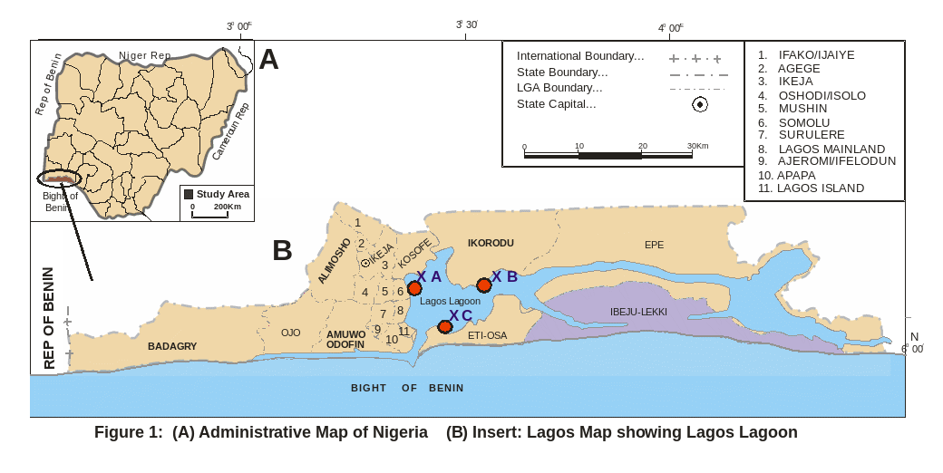
Lagos lagoon (Figure. 1), Nigeria is located on longitude 3020′ and 3050′W and latitude between 6024′ and 6036′N. It is the largest of lagoon systems in West African coast covering 208 Km2. It is an open tidal estuary. It is fed in the north by Ogun River, which is the major source of water to the lagoon; the southern margin is bounded by Five Cowries Creeks. The adjacent Lekki and Epe lagoons bounded it in the east. The Badagry creek is the main waterway leading to Badagry and Nigeria/Benin Republic border. It opens into the Atlantic Ocean via the Lagos habour. Connected with Five Cowries Creek is Kuramo water, which is a channel, which it runs through the mangrove swamps. Previously reviews include several studies on benthic, fishes and fisheries of this water body (Olaniyan, 1957; Oyenekan, 1972; Fagade and Olaniyan, 1972, 1973; Yoloye, 1976; Kusemiju et al. 1983; Ajao, 1990; Brown, 1991; Lawson, 1991, 1998).
2.2 Collection of specimens:
2,167 specimens of mudskipper, Periophthalmus papilio were caught between May 2006 and May 2008 with non-return valve traps from mud flats of the mangrove swamps of Lagos lagoon Services of local fishermen were employed in setting up traps and diurnal collections of the fish. They were preserved in 4% formaldehyde prior laboratory procedures.
2.3 Laboratory procedures and data collection:
Biometric data on total length (TL) to the nearest 1 mm and body weight measurements (BW) to the nearest 0.01g were recorded. Further laboratory analysis was carried out by opening bellies of the specimens to ascertain sex and gonad weight (GW). The maturity stages were determined by naked eye and microscopic examinations of the gonads. Methods of classifications of gonadal stages followed those of Jone (1974), Blay and Eyeson (1982), Marcus (1982), and Ugwumba (1984). Sex ratio was determined by counting numbers of male and female specimens. The ratios were tested with Chi-squared analyses (X2). The annual gonadal cycle was determined from the changes in gonad weight as indicated by the gonadosomatic index (GSI). The GSI was estimated from the relationship:
GSI = GW . 100 . BW-1.
The relationship between GW and BW of the fish was obtained from equation:
GW=a+bBW. Parameters a and b were estimated from the least squares regression method.
Egg size or oocyte diameter (OD) was measured with a graduated micrometer that was mounted in the eyepiece of a binocular microscope. The regression coefficient, r was calculated to the nearest 0.1 mm. The relationship that existed between OD and fish TL was determined from equation:
OD=a+bTL
3.0 Results
3.1 Sex ratio:
In the current study a total of 2167 specimens of P. papilio were collected from the mangrove swamps of Lagos lagoon. This number comprised 281 immature, 779 male and 1107 female individuals giving a sex ratio of 1male:1.42 females. A Chi square (X2) revealed a significant departure(X2=57.04>X21, 0.05=3.84), from 1male:1female sex ratio indicating females were significantly more than males.
3.2 Gonadosomatic index:
In the present study, GSI varied with sex (Table 1). It varied in between 0.01 and 0.48% (0.104±0.004%) and from 0.11 to 8.40% (2.69±0.003%) in females.
Table
1. Summary of the GSI in male and female P.
papilio in Lagos lagoon. Sex Range
(%) Mean
±S.E (%) Min Max. Males
0.01 0.48 0.104±0.004 Females
0.11 8.40 2.69±0.03
The gonad weight-total length relationships in P. papilio are expressed as:
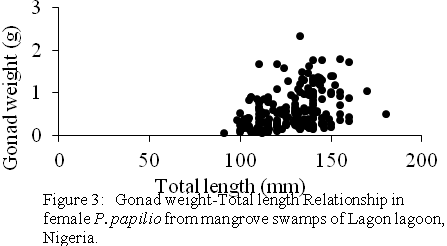
For males (Figure 2)
GW=0.0434+0.0005TL(n=126,r=0.4385)
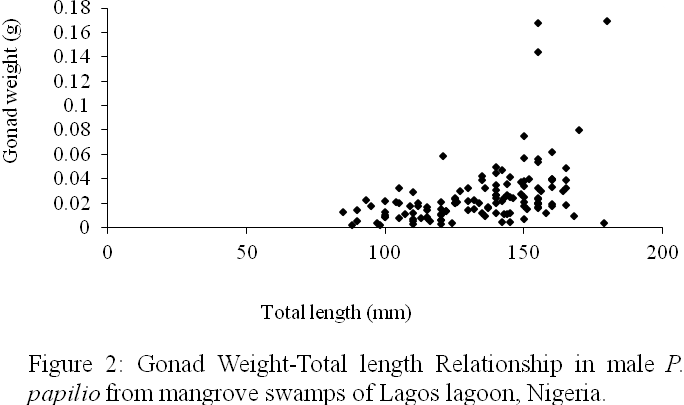
For females (Figure 3)
GW=0.7502+0.0106TL(n=199,r=0.3835)
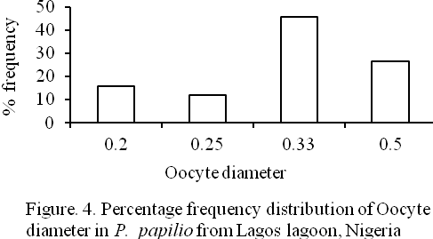
3.3 Oocyte diameter:
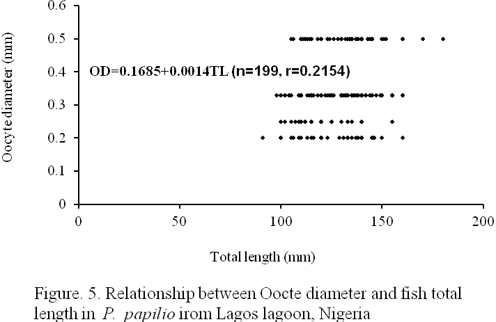
Four (4) different egg sizes were observed in the species viz: 0.2., 0.25, 0.33, and 0.50mm oocyte diameters representing 15.56, 12.06, 45.73, and 26.63% of the total eggs respectively (Figure 4). The mean egg diameter was 0.36±0.002 mm. The relationship between oocyte diameter (OD) and fish total length (TL) as presented in Figure 5 is:
OD=0.1685+0.0014TL (n=199, r=0.2154).
3.4 Stages of maturity: Seven (7) maturity stages were encountered. The macroscopic and microscopic descriptions of the maturity stages for the species are presented in Table 2.
.
Table 2: Stages of maturity in P. papilio from mangrove of Lagos lagoon, Lagos, Nigeria
|
Maturity stages |
Males |
Females |
|
Stage I (Immature) |
The abdominal cavity. Macroscopic examination did not reveal presence of accessory sexual organs, which were associated with males and females. The microscopic examination showed no sex differentiation. The cells associated with the gonads were rudimentarily developed and could not be differentiated. Hence the specimens were classified as immature. |
|
|
Stage II (Immature and Developing)
|
Macroscopically, the testes were flattened, 1-2mm broad, whitish and lobed. They occupied 1% of the body cavity and the accessory sexual organs were visible. Microscopically, the testicular wall was thick with primary spermatocytes predominating periotoneum. The mesothelium of the peritoneum was very thick. The stoma and interlobular septa were very conspicuous. |
External or macroscopic appearance of the ovaries showed that they were small and rounded with a rough surface and soft texture. They were creamy in colour and measured between 1/8th (12.5%) and 1/4th (25.0%) of the length of the abdominal cavity. They were translucent with blood vessels forming internally; none of the oocytes were visible through the ovary wall. Histological appearance was characterized by the presence of many oocytes (0.025-0.05mm). The larger oocytes had cytoplasm vacuoles. The ovary wall was thick at 50µm and folded. The oocytes were irregularly shaped but few were rounded. |
|
Stage III (Ripening)
|
In early stage (Phase 1) of ripening, the testes became fatter, off white and occupied 1/8th of the abdominal cavity. Blood vessels were visible through testis wall. Gonad length: width ratio was 2.8. Primary spermatocytes and spermatids were pronounced. In the late stage (Phase 2) of the ripening, the testes became firm and whiter and occupied 1/5th of the abdominal cavity. Lobulation of the right and left testes started. The length: width ratio was 2.4. The accessory sexual organs were of equal length with the testes. The secondary spermatozoa were present. The septa were not fully organized. |
The ovaries were swollen and lobed when observed macroscopically. A heavy network of vessels appeared externally on the surface of the ovary wall. Yellowish oocytes were visible to naked eye through the ovary wall. The gonad extended for about 60 – 70% of the abdominal cavity. The histological observation of the ovaries at this stage showed that many oocytes of between 0.1 and 0.2mm were present. Larger oocytes with cytoplasmic vacuoles were very few and had small yolk droplets. The primary and secondary vitellogenic oocytes dominated while tertiary vitellogenic oocytes were represented in the gonad. The cytoplasm of larger oocytes was filled with densely staining yolk granules. The ovarian wall was 70µm thick. |
|
Stage IV (Ripe) |
The testes were fully swollen and multilobed at this stage but did not occupy more than 1/4th of the body cavity. The colour was creamy white. The accessory sexual organs grew past the testes. The secondary and tertiary spermatocytes were dominant while few primary spermatocytes were represented. The testicular wall was 30µm. The testicular septa were well organized and distinct.
|
External appearance showed that the ovaries at this stage were almost filling the body cavity occupying 80 –90% of abdominal cavity. The shedding of eggs has not commenced and otherwise soft. The eggs were rounded with a rough granular surface given a hollow sac like appearance. Blood vessels coalesced to form larger ones on the external surface of the ovary wall. Yellowish colour was possible due to the large yellow oocytes that were visible through ovary wall. The histological observation of the gonads showed that the secondary and tertiary vitellogenic oocytes dominated the gonad with very few primary oocytes. The theca externa were prominent. The hyaline oocytes were present but usually collapsed by histological processing. Ovary wall was 90µm thick; many oocytes were between 0.2 to 0.5mm in diameter but usually 0.35mm in size. Many oocytes were at stages II and III. There were blood vessels internally but some of the yolky oocytes were atretic. |
|
Stage V (Ripe running) |
The testes were broadest, mostly firm but some were flaccid and highly lobulated. They were completely white but the posterior tip sometimes grew with speckled appearance. The accessory sexual organs were fully developed and became longer than the testes. No blood vessels and thick milt exuded on slight pressure before preservation. Testis length: width ratio was 2.2 and it extended for 50% of the abdominal cavity. The lumen contained spermatozoa. The empty spaces in the lumen also contained spermatocytes and spermatozoa. Most of the spermatozoa migrated towards the periphery of the lobule and the more advanced stages such as primary and secondary spermatocytes and the spermatids were found towards the interior. The testicular wall reached 30µm |
The oocytes or eggs flowed from the vent on slight pressure and the ovary occupied 99% of the abdominal cavity and rendered alimentary canal and gut almost inconspicuous. Oocytes looked exactly like those in the ripe stage and were laid singly with space (septa) in between as shown in most of the oocytes were in their tertiary vitellogenic stage. |
|
Stage VI (Spent) |
The testes reduced in size and sometimes very small, flaccid and walls were hard in texture. They were dark brown colour and no blood vessel visible. Milts were absent, testis length: width ratio was 3.2 and gonad extended for 30% of the abdominal cavity. The testis had unfilled lumen with inactive spermatozoa. The testicular wall reached 40µm. The accessory sexual organs well developed and longer than what obtained in stage V. The septa disappeared and mesothelium was thickest.
|
The ovaries were reduced in size, flaccid but ovary wall was touch and smooth with no granulation. The colour was dark red. The residual oocytes were present and visible through flabby wall. The ovary length: width ratio was 4.5 and gonad extended for 50% of the abdominal cavity. The diameters of the oocytes were very few and had a thin densely staining cytoplasm. A few atretic residual oocytes were present and invasion of oocytes by follicular cells was noted. There was a dense network of blood vessels, which indicated a high level of oocyte atresia. The septum was disorganized and there was no empty follicular coat. The ovary wall was 300µm thick. The lumen of the ovary contained much debris that was residual cells. |
|
Stage VII (Resting and Recovering)
|
Dark patches visible through the testis wall. The testes were about a third of length of body cavity or less and firmer than what obtained at spent stage and 3mm broad. A big cavity was seen at the center of the gonad and residual spermatozoa were present at the lumen. The mesothelium of the peritoneum thickened.
|
Externally, ovaries were firmer than spent stage but mainly red in colour. It occupied 60% of the body cavity and none of the residual oocytes were visible through the ovary wall. The residual atretic oocytes were present when observed microscopically but the septum was not very organized. Reorganization of ovigerous lamellae started. A few reabsorbing oocytes were sometimes present. |
|
|
|
|
|
|
|
|
|
|
|
|
|
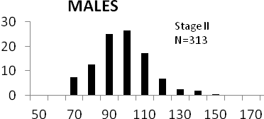
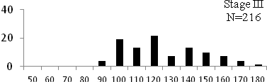
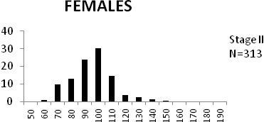
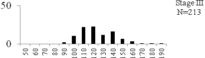
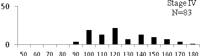
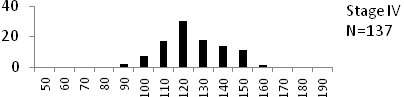
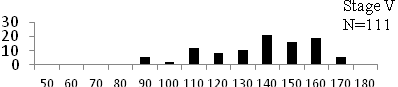
Total length (mm) Total length (mm) Figure
6. Length frequency distributions of P.papilio at
different maturity stages (II – VII) in the mangrove
swamps of Lagos Lagoon Nigeria |
|

3.5 Size at maturity:
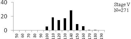
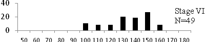
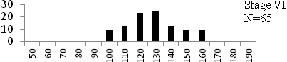
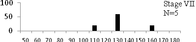
Length frequency histograms of the species at different stages of maturity are presented in Figure 6. Fish on stage II measured between 70 and 150mm TL for males and females respectively. The modal length for 26.52 of males and 30.44% of females was 100mm TL. Those on stage III were between 80 to 160 for males and 90-190mmTL for females, the modal length was 120mm. 25.0 and 23.01% of the males and females respectively were in this modal length. Stage IV fish ranged from 90 to 180 mm TL (for males) and 90-160 mm TL (for females), the modal lengths were 140mm for 17.53 males and 120 mm TL for 23.53% females. 20.72% of the males were at ripe stage (V) between 90 and 160 mm TL and from 90-160mm for females. The modal lengths were 140 and 120mm TL respectively. At 120 mm TL, most females were already at ripe (IV) and ripe running (VI) stages earlier than the males. The fish in stage VI ranged from 100 -160 mm TL. In Stage VII, 60% of the specimens were 130mm TL. The females were between 110 and 160mm TL, 28.57% of them measured 120mm TL.
4. Discussion
In the present study on the fish Periophthalmus papilio, sex ratio, gonadosomatic index, egg diameter and stages of maturity were discussed in order to clarify some characteristics of its reproductive biology. Sex ratio of this species in the mangrove swamps of Lagos lagoon indicated that females were significantly more than the males giving a sex ratio of 1 male: 1.42 females. This is a departure from theoretical 1male to 1female sex ratio. High percentages sex ratio in favour of males during the spawning period was reported in non related species like Elops lacerta (Ugwumba, 1984; Lawson and Aguda, 2010) and Chrysichthys walkeri (Kusemiju, 1978) and while ratios in the favour of latter were reported in Ethmalosa fimbriata (Fagade and Olaniyan, 1972; Blay and Eyeson, 1982) and Mugil cephalus (Lawson 1991) in some West African lagoons
The eggs of this species were as small as 0.2 and as big as 0.50mm (mean= 0.36±0.002mm) in the lagoon (Figure 4). The prominent oocytes were those with 0.30 mm in diameter, least were 0.25mm egg diameters. This was in supports of the reports of Jone (1974) on Scophthalmus maximum and Marcus (1982) on Ilisha africana. The egg size in teleosts varies with the size of female (Thorpe et al 1984), with food supply (Bagenal, 1969) and with species (Bagenal, 1969; Hay and Brett, 1988). The gonadosomatic index of the fish varied between 0.01 and 0.48 in males and from 0.11 to 8.40% in females. Females showed higher GSI values in Table 1. This was an indication that less than 0.48 in males and 8.40% of female body mass were committed to gonad development by this species. The GSI had been used to describe the development of gonads in Pike, Esox lucius (Danilenko, 1983). GSI increases progressively with increased percentage of the ripe individuals towards the spawning seasons (Mohamed 2010). The most common practice for determination of determination of a species spawning season is the establishment of its GSI and the histological examination of the gonads (El-Greisy, 2000; Assem, 2000 and 2003; Honji et al. 2006).Gonad weight and total length measurements were positively correlated in males (r=0.439, Figure 2) and females (r=0.384, Figure 3).
In this study seven stages of gonad development were observed (Table 2). These stages were classified as:
Pre-spawning class which was represented by immature, immature and developing, ripening stages.
Spawning represented by ripe and ripe running stages and
Post-spawning class which was represented by spent and recovering/resting periods of the fish in Lagos lagoon.
Observations of these stages of maturation in the fish though with modifications were in conformity with that of most teleosts (Assem, 2002 and 2003). The presence of spermatocytes and oocytes at different stages of development was an indication that this species belongs to the fish with prolonged and fractional spawning season. Therefore, the fish may spawn more than once during the spawning season. This was in agreement with reports of Salem et al (1994) on Mugil seheli, El-Greisy (2000) on Diplodus sarus, Honji et al (2006) on Merluccius hubbsi, Garcia Diaz et al (2006) on blacktail comber, Serranus atricauda, and Mohamed (2010) on Merluccius merluccius. Maturation process in P. papilio was similar to that of its relative, Boleophthalmus pectinirostris in Midori River, Japan (Washio et al 1991). Thus maturation proceeds in this species as follows: In females, a portion of the oocytes in immature ovaries develop to the secondary yolk stage and form an oocyte group. Then parts of this oocyte develop further in turn to be spawned, leaving the remainder of the oocyte group at the secondary yolk stage. At repetition of this process, spawning comes to an end with the declination of oocyte development in the ovary. In males, the spermatids develop to primary spermatocytes which further divide to secondary spermatocytes which to be spawned as spermatozoa. Like in females, repetition process makes spawning to come to an end with declination of milt development in the testis.
Conclusion
The current study on mudskippers, P. papilio in Lagos lagoon, Nigeria showed a record of higher number of females than males (sex ratio of 1male:1.42 females). This was significantly different but a departure from the theoretical 1male:1female ratio. The smallest egg measured 0.20 in diameter while the biggest was 0.50 mm (mean=0.36±0.002 mm). There are seven classes of maturity representing the pre-, spawning and post spawning periods of the fish in Lagos lagoon. At 90 mm TL both sexes attained ripe stage, spent between 100-160mm TL. A study is currently in progress to investigate the diet composition, fecundity, taxonomy, age and growth patterns of this commercially valued fish species in Lagos lagoon and other water bodies in Lagos. Therefore data from this work will be useful as baseline data for carrying out further ecological and biological studies for proper management and conservation of fisheries resources of Lagos lagoon.
Acknowledgements.
Author acknowledges both Departments of Fisheries of Lagos State University and University of Lagos, Nigeria for the use of their laboratories for this study. Also acknowledge is Mr. Daniel James for secretariat assistance.
Corresponding Author:
Dr. LAWSON Emmanuel Olugbenga
Department of Fisheries
Lagos State University, Lagos, Nigeria.
E-mail: [email protected].
References
1 Irvine FR. The Fishes and Fisheries of Gold coast. Crown Agent: London. 1947; 352pp.
2. Food and Agricultural Organization (FAO). Field guide to commercial marine Resources of the Gulf of Guinea. FAO/UN Rome (Italy), 1990; 265pp
3. Ip YK, Low WP, Lim ALL, Chew SF. Changes in lactate content in the gill of the mudskippers, Periophthalmus chrysospilos and Boleophthalmus boddaerti in response to environmental hypoxia. Journal Fish Biology 1990; 36: 481-487.
4. Lawson EO. Distribution patterns, age determination and growth studies of Mudskipper, Periophthalmus papilio in mangrove swamps of Lagos lagoon, Lagos, Nigeria. Journal of Research and Review in Science 2004a; 3: 293-297.
5. Murdy EO. "A Taxonomic Revision and Cladistic Analysis of the Oxudercine Gobies (Gobiidae: Oxudercinae)". Records of the Australian Museum Suppl. 1989; N°11: 1–93.
6. Etim L, King RP, Udo MT. Breeding, growth, mortality and yield of the mudskipper, Periophthalmus barbarus (Linneaus 1766) (Teleostei: Gobiidae) in the Imo River estuary, Nigeria. Fisheries Research 2002; 56 (3), 227-238.
7. Jaafar Z, Larson HL. A new species of mudskipper, Periophthalmus takita (Teleostei: Gobiidae: Oxudercinae), from Australia, with a key to the genus. Zoological Science 2002; 25 (9): 946–952. doi:10.2108/zsj.25.946. PMID 19267605.
8. Khaironizam MZ, Norma-Rashid Y. Length-weight relationship of mudskippers (Gobiidae: Oxudercinae) in the coastal areas of Selangor, Malaysia. NAGA Worldfish Cent Quart 2002; 3: 20–22.
9. Udo MT. Intersexual plasticity in aspects of the biology of the mudskipper Periophthalmus barbarus (Gobiidae) in the mangrove swamps of IMO Estuary, Nigeria. J Environ Sci. 2002; 14(1), 95-101.
10. Lawson EO. Food and feeding habits of mudskipper, Periophthalmus papilio in mangrove swamps of Lagos lagoon, Lagos, Nigeria. Journal of Research and Review in Science 2004b; 3: 355-358.
11. Swanson BO, Alice CG. Kinematics of aquatic and terrestrial escape responses in mudskippers. Journal of Experimental Biology 2004; 207, 4037-4044. doi:10.1242/jeb.01237
12 Sarimin, AS, Ghaffar MA, Mohamed CAR. Variation of Ca, Sr, Ba and Mg in the Otolith of Mudskipper in West Coast of Peninsular Malaysia. Pakistan Journal of Biological Sciences 2009; 12(3): 231-238. ISSN 10288880
13. Jaafar Z, Lim KKP, Chou LM. Taxonomical and Morphological Notes on Two Species of Mudskippers, Periophthalmus walailakae and Periophthalmodon schlosseri (Teleostei: Gobiidae) from Singapore. Zoological Science 2006; 23(11): 1043-1047. 2006 doi: 10.2108/zsj.23.1043
14 Olaniyan CIO. The seasonal variation in plankton in the Lagos Habour, Nigeria. Ph.D Thesis, London University. 1957; 176pp
15 Oyenekan JA. A survey of the Lagos lagoon benthos with particular reference to molluscs. M.Sc dissertation, University of Lagos. 1972; 220pp.
16 Fagade SO, Olaniyan CIO. The biology of the West Africa Shad, Ethmalosa fimbriata (Bowdich) in the Lagos lagoon, Nigeria. Journal Fish Biology 1972; 4: 519-534.
17 Fagade SO, Olaniyan CIO. The food and feeding interrelationships of the fishes in Lagos lagoon, Nigeria. Journal Fish Biology 1973; 5: 205-225.
18. Yoloye V. 1976. The Ecology of the West African bloody Cockle, Anadara (senilis) senilis (L). Bull. d’l.F.A.N.T., 38 Ser. 1976, A(1): 25-55.
19 Kusemiju K, Oki AA, Graham-Douglas M. On the biology of an estuarine population of the Clupeid, Pellonula afzeliusi in Lagos Lagoon, Nigeria. Hydrobiology 1983; 102: 55-59.
20 Ajao EA. The influence of domestic and industrial effluents on populations of sessile and benthic organisms in Lagos lagoon. Ph.D Thesis, University of Ibadan, 1990; 413pp.
21 Brown CA. Community structure and secondary production of benthic macrofauna of Lagos lagoon harbour. M. Phil dissertation, University of Lagos, Nigeria. 1991; 359pp.
22. Lawson EO. Biology of the Grey mullet, Mugil cephalus, L. in Lagos lagoon, Nigeria. M.Sc dissertation, University of Lagos, Nigeria. 1991; 88pp.
23. Lawson EO. Bio-ecology of the Mudskipper, Periophthalmus papilio in the mangrove swamps of Lagos lagoon, Nigeria. Ph.D. Thesis, University of Lagos, Nigeria, 1998; 180pp.
24. Jone A. Sexual maturity, fecundity, and growth of Turbot, Scophthalmus maximum (L). Journal of Marine Biology Association of United Kingdom 1974; 54: 109-125.
25 Blay J Jr, Eyeson KW. Observations on the reproductive biology of the Shad, Ethmalosa fimbriata (Bowdich) in the coastal waters of Cape coast, Ghana. Journal Fish Biology 1982; 21: 485-496.
26 Marcus O. The biology of the Clupeid, Ilisha africana (Bloch) off the Nigerian coast. Ph.D Thesis, University of Lagos, Nigeria. 1982; 278pp.
27 Ugwumba OA. The biology of the ten pounder, Elop lacerta (Val.) in the freshwater, estuarine and marine environment. Ph.D Thesis, University of Lagos, Nigeria. 1984.
28 Lawson EO, Aguda AF. Growth patterns, diet composition and reproduction in the ten pounder, Elops lacerta from Ologe lagoon, Lagos, Nigeria. Agric. Biol. J. N. Am., 2010; 1(5): 974-984 doi:10.5251/abjna.2010.1.5.974.984.
29. Kusemiju K. Distribution, reproduction and growth of catfish, Chrysichthys walkeri (Gunther) in the Lekki lagoon, Nigeria. Journal Fish Biology 1976; 8: 453-458.
30 Thorpe JE, Miles MS, Keay DS. Developmental rate, fecundity and egg size in Atlantic salmon, Salmo salar L. Aquaculture 1984; 43: 289-305.
31 Bagenal TB. The relationship between food supply and fecundity in brown trout, Salmon trout, Salmo trutta. Journal Fish Biology 1969; 1: 169-82.
32 Hay DE, Brett BR. Maturation and fecundity of Pacific herring (Clupea harengus pallasii), an experimental study with comparisons to natural populations. Can. Journal Fish Aquatic. Science 1988; 45: 399-406.
33 Danilenko TP. The reproductive cycle of the Pike, Esox lucius, L in the Kanev Reservoir. Hydrobiology 1983; 18(4): 21-27.
35 Assem SS. The reproductive biology and histological characteristics of pelagic Carangid female Caranx crysos, from the Egyptian Mediterranean sea. J. Egypt. Ger. Scc. Zool. 2000 31(C ) : 195-215.
36 Assem SS. The reproductive biology and histological and ultrastructural characteristics of the ovary female pelagic fish Pagellus erythrinus from the Egyptian Mediterranean water. J. Egypt. Ger. Scc. Zool. 2003; 42: 77-103.
37 Honji RM, Vas-dos-Santos AM, Rossi WS. 2006. Identification of the stages of ovarian maturation of the Argentine hak Merluccius hubbsi, Marini, 1993 (Teleostei: Merlucciidae) advantages and disadvantages of the use of the macroscopic and microscopic scales. Neotrop. Ichthiol. 2006; 443: 329-337.
38. Assem SS. The reproductive biology and histological characteristics of pelagic carangid females Caranx crysos, from the Egyptian Mediterranean sea. J. Egypt. Ger. Soc. Zool. 2002; 42: 77-103.
39 Salem SB, Zaki MI, El-Gharabawy MM, El –Shorbagy IK, El-Boray KF. Seasonal histological changes in the ovaries of Mugil seheli from Suez Bay. Bull. Nat. Inst. Oceanogr. Fish. ARE, 1994; 20 (1): 235-249.
40 Garcia Diaz M, Gonzalez JA, Lorente MJ, Tuset VM. Spawning season, maturity sizes, and fecundity in blacktail comber (Serranus atricauda) (Serranidae) from the eastern-central Atlantic. Fish Bull., 2006; 104:159-166.
41 Mohammed A.A. The reproductive biology and the histological and ultrastructural characteristics in ovaries of the female gadiidae fish Merluccius merluccius from the Egyptian Mediterranean water. African Journal of Biotechnology 2010; 9(17): 2544-2559.
42. Washio M, Tsutsui M, Takita T. Age and growth of mudskipper Boleophthalmus pectinirosiris distributed in the mud flat of the Midori River, Kumamoto Prefecture. Nippon Sulsan Gakkaishi,1991; 57, 637-644
9/30/10
ISTS 2002M01 MISSION AND SCIENCE OF MUNIN
NEBRASKA’S HEALTH SCIENCE CENTER OFFICE OF REGULATORY AFFAIRS
PGCE PRIMARY SCIENCE SPECIALIST PATHWAY 20182019
Tags: 3(11) aspects, 3(11), aspects, science, journal
- OBECNĚ ZÁVAZNÁ VYHLÁŠKA OBCE ČKYNĚ Č 12015 O STANOVENÍ
- NYT FRA BRUGERGRUPPEN – MARTS 2021 DET SENESTE ÅR
- B21 WRITTEN MEDIATION TASK WRITE AN EMAIL TO YOUR
- UWAGI I PROPOZYCJE ZMIAN DO PODSTAWOWYCH ZASAD GOSPODARKI FINANSOWEJ
- ROOM RESERVATION CONFIRMATION EASTERN KENTUCKY UNIVERSITY OFFICE OF STUDENT
- TAMMSFLEX NS & TAMMSFLEX SL NSF APPROVED TWO PART
- 6 LAW OF THE AZERBAIJAN REPUBLIC ON WATER ECONOMY
- A) DIEGO HABLA DE SU PADRE COMPLETE ESTE PÁRRAFO
- PRISPEVKI ZA SOCIALNO VARNOST BROŠURA O PLAČEVANJU PRISPEVKOV ZA
- WRITING A MYTH A MYTH HAS BEEN DEFINED AS
- PREFEITURA DE CONCEIÇÃO DA BARRA ESTADO DO ESPÍRITO SANTO
- I WNIOSEK DO PRZEWODNICZĄCEGO KOMISJI KOMÓRKA ORGANIZACYJNA …………………………………………………………………………………………………………………………………………… WNIOSKUJĘ
- HARMONOGRAM PRZEBIEGU POSTĘPOWANIA HABILITACYJNEGO DR ANNY MARII JESZKI PRZEPROWADZANEGO
- A YEAR AT A GLANCE TAYLOR SCHMIDT SUMMER SCHOOL
- DYRECTO EN BRUSELAS ¿CÓMO APOYA LA UNIÓN EUROPEA LOS
- SERVICIO DE PREVENCIÓN SERVICIO DE PREVENCIÓN AJENO RIESGOS GENERALES
- C OMPILE PROGRAM DETAILS WORK SHEET PROGRAM OR COALITION
- G REENVILLE COUNTY BUILDING SAFETY DIVISION GREENVILLE COUNTY SQUARE
- POWERPLUSWATERMARKOBJECT3 CONVOCATORIA DE SUBVENCIONES PARA LA FINANCIACIÓN DE PROGRAMAS
- ZAŁĄCZNIK NR 2 DO WZORU UMOWY (ZAŁĄCZNIK NR 12
- CONTRATOS MIGUEL SERVET MEMORIA CONTRATO Y LÍNEA
- RAPPORTNR 6 2014 FRA HESTKINN NATURBEITEMARK MED ORKIDEER 2562013
- 1 SOFTWARE DE LA PRÁCTICA ADEMÁS DEL STATGRAPHICS
- KAKO SE NOSITI SA ANKSIOZNOŠĆU I STRESOM ZA VRIJEME
- RESOLUCION EXENTA SII N°79 DEL 19 DE JULIO DEL
- Č ÍNA (ČÍNSKÁ LIDOVÁ REPUBLIKA – ČLR) HLM PEKING
- KAIROS – EDAD MODERNA LA EUROPA DEL BARROCO PROYECTO
- SPN 132 SPRING 2005 PRACTICE IMPERFECT TENSE (AR ER
- LOS JÓVENES QUIÉNES HACEN LA REVOLUCIÓN QUIÉNES SON LOS
- SUBSKILL MULTIPLES AND FACTORS I FACTOR TO SOLVE
 GUIDE SPECIFICATION SECTION 28 23 29 ROADWAY VIEWING SOLUTION
 Notice-of-Appointment-of-Election-Agents-Hillside
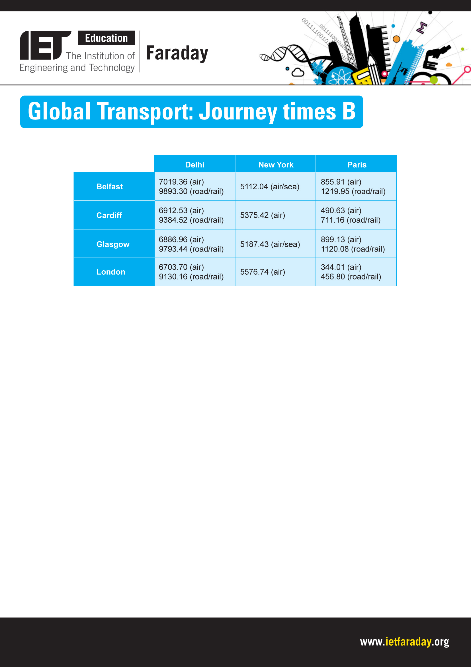 THE TABLE ABOVE SHOWS THE DISTANCES BETWEEN CITIES IN
NR POSTĘPOWANIA ZP42021ZO TRZEBNICA 03032021 R ZAPROSZENIE DO SKŁADANIA
PRACA NA ZAJĘCIACH 1 PRAWO RODZINNE – SPRAWY MAŁŻEŃSKIE
EK SERMAYE PİYASASI KURULU KURUMSAL YÖNETİM İLKELERİ GİRİŞ ÖNEMLI
A KÖZÉRDEKŰ ADATOK MEGISMERÉSÉRE IRÁNYULÓ IGÉNYEK BENYÚJTÁSÁNAK ÉS INTÉZÉSÉNEK
 POR LA PRESENTE AUTORIZO A LA ASOCIACIÓN ÁGORA ALTAS
 MEDIATION VERSUS MODERATION TABLE OF CONTENTS 1 AN OVERVIEW
 MAGISTRÁT MĚSTA OLOMOUCE ODBOR STAVEBNÍ ODD STÁTNÍ SPRÁVY NA
 BASE CONSOLIDADA PODER LEGISLATIVO NOTAS A LOS ESTADOS FINANCIEROS
USING NIH COMMONS TO SUBMIT YOUR 2590 PROGRESS REPORT
 FUNDAMENTOS DEL DERECHO TEMA 5 SUJETOS DE DERECHO Y
KIT DEMANDE DAIDE JURIDICTIONNELLE POUR CONTESTER UNE OQTF (OBLIGATION
MLA CONVENTIONS FOR USING PARENTHETICAL CITATIONS WORKSHEET USE PARENTHESES
LEY PARA LA COORDINACION DE LA EDUCACION SUPERIOR TEXTO
 1 SUTRUMPINTAS DĖSTYTOJO PRISTATYMAS VARDAS PAVARDĖ AISTĖ LAZAUSKIENĖ MOKSLINIS
 AFTER THE REFERENDUM CONSTITUTIONAL CHANGE IN THE EVENT OF
Informatikai Rendszerek Számítógépes Vállalatirányítás 1 Mutassa be a
SOUTHNORTH DEVELOPMENT MONITOR (SUNS) 6272 FRIDAY 15 JUNE 2007
