LOOKING AT THE HEART THE PURPOSE OF THIS PROCEDURE
28 FISCAL POLICY IN KENYA LOOKING TOWARD THE MEDIUMTO6 I F ONE WERE LOOKING FOR A MAN
61 REPORT OF THE WORKING GROUP LOOKING AT THE
ABSTRACT SECOND ORDER COHERENCE A NEW WAY OF LOOKING
ARE YOU LOOKING TO VOLUNTEER? DO YOU HAVE A
ATELIER TEN IS LOOKING FOR AN ENERGY MODELER WITH
Title

Looking at the heart
The purpose of this procedure is:
to investigate the general structure of the heart
to appreciate the differences between the muscles in different parts of the heart, and the structures such as valves and tendons that make the heart work
to appreciate the differences in the structures of the blood vessels associated with the heart.
Procedure
SAFETY: Wear eye protection whenever there is a risk to the eyes, for example, when changing scalpel blades, cutting cartilage or if the heart you are working with has been preserved.
Clean the work area carefully after the investigation.
Investigation 1 – Looking at the outside of a heart
Note the general shape and size of the heart.
Measure its size and mass. Estimate its external volume.
Identify the vessels entering and leaving the heart. Arteries have thick, rubbery walls. Veins have much thinner walls. Feel inside these vessels with your fingers and feel the texture and strength of both vessels. Try to describe what you feel.
Look inside the main arteries and veins. If you see any structures attached to the walls, try to decide what they are, what they might do and how they might work.
Identify the atria and ventricles. Try to describe any differences in the structures of the walls of the atria and ventricles.
Decide which you think is the right side of the heart and which is the left. What has helped you to decide?
Examine the surface of the heart for blood vessels.
Note the colour and texture of the different parts of the heart.
Investigation 2 – The internal structure of the heart

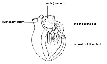
Diagram 1 Diagram 2
Look at the first diagram. Make a long cut down through the aorta and the left ventricle to the tip of the heart (‘apex’), as shown in the diagram. The position of the blood vessels on the surface will help you to make this cut in the right place.
Pull the edges of the ventricle apart and examine the inside of the ventricle and the aorta. At the base of the aorta, you will see the structures mentioned in d. Examine them carefully.
Look at the second diagram. Cut upwards carefully into the left atrium along the line shown in the diagram.
Measure and record the thickness of the walls of the atrium and the ventricle.
Examine the right side of the heart in a similar way.
Look at the areas where an atrium joins a ventricle. Examine the structures there. These are valves separating the chambers of the heart. You should see flaps of thin tissue, with tough ‘threads’ attached to the base of the flaps. Count how many threads there are on each side of the heart. Think about how these valves might work.
Record sheet for student observations
The Heart
Overall size:
Mass:
Estimated external volume:
|
Vessel type 1
|
Vessel type 2 |
|
Atrium
Thickness of wall: |
Ventricle
Thickness of wall: |
|
Valve type 1
Position: Way of working: |
Valve type 2
Position: Way of working: |
©
Nuffield Foundation / Biosciences Federation 2008 •
Downloaded from Practicalbiology.org • PAGE
BANKING STRUCTURE IN INDIA LOOKING AHEAD BY LOOKING BACK1
BECHA IS LOOKING FOR A NEW BOARD MEMBER BEXLEY
BRISTOL MIND IS LOOKING TO EMPLOY TWO EXCEPTIONAL PEOPLE
Tags: heart the, the heart, heart, looking, procedure, purpose
- SABOR DE AMOR – DANZA INVISIBLE CONTENIDOS CONTENIDOS
- 3GPP TSGRAN WG4 98BISE R42104971 ELECTRONIC MEETING 12TH –
- CLUB EVENT CHECKLIST HOLD A MEETING WITH CLUB
- 15 LICENSE AGREEMENT THIS AGREEMENT IS EFFECTIVE ON
- LALOR GARDENS PRIMARY SCHOOL NO 5532 134 KINGSWAY DRIVE
- ATTACHMENT 3 1 1 REPRESENTATIVE OF THE RCUH THE
- EKER – BYK TROFESİ 2014 BURGAZ KUPASI YELKENLİ YAT
- (MODELO DE FACTURA EMITIDA POR ASOCIACIONES SIN ANIMO DE
- TEORÍA ANTROPOLÓGICA CONTEMPORÁNEA PROGRAMA DE POSGRADO EN ANTROPOLOGÍA SOCIAL
- SPECIALTY SPECIFIC REQUIREMENTS FOR APPLICATION IN BEHAVIORAL & COGNITIVE
- FORMULARE ȘI MODELE CONŢINE FORMULARELE DESTINATE PE DE
- FROM DOMAGOJ SAJTER [MAILTODOMAGOJSAJTERGMAILCOM] SENT TUESDAY FEBRUARY 03 2015
- HOSPITAL MARIA AUXILIADORA DECENIO DE LAS PERSONAS CON DISCAPACIDAD
- WYMAGANIA EDUKACYJNE KLASA II GIMNAZJUM HISTORIA I NA OCENĘ
- PIPER THE PIG HAS A DAY OUT BY HEATHER
- RADA® SOLUTIONS FOR CENTRAL RECIRCULATION SYSTEM CONTROL RADA EMC
- NO 32 VOL1 THN XVI NOVEMBER 2009 ISSN 08548471
- ATTACHMENT II FINANCIAL AND COMPLIANCE AUDIT ATTACHMENT THE ADMINISTRATION
- NORMAS DE USO DE LA PISTA DE ATLETISMO
- 11 III ODŮVODNĚNÍ OBECNÁ ČÁST A ZÁVĚREČNÁ ZPRÁVA O
- OBČINA POSTOJNA 7 SEJA OBČINSKEGA SVETA GRADIVO K TOČKI
- ATTACHMENT G SPECIFIC WEBSITE CHARACTERISTICS ADMINISTRATIVEGENERAL PRIVACY STATEMENT
- NOTIFICAION IN EXERCISE OF THE POWERS CONFERRED BY CLAUSE
- BÜRO VE DESTEK HİZMETLER İŞ AKIŞI DOKÜMAN NO İLK
- LEGIÓN DE MARÍA ¿QUÉ ES Y CUÁL ES SU
- ATTACHMENT 1 RELATIONS WITH OTHER PROGRAMS DATE WE ARE
- TEMA 15 ESTRATEGIAS DE PRUEBA DEL SOFTWARE
- 2 ANEXO 1 FORMATO DE RESOLUCIÓN ADMINISTRATIVA INSTITUCIONAL PARA
- ATTACHMENT 1 SITE PLAN FOR OC EXPANSION
- POWERPLUSWATERMARKOBJECT3 PHYSICAL EDUCATION YEAR PLAN FOR THE YEAR OF
EJERCICIOS DE DIVISIÓN SILÁBICA LA SIGUIENTE FRASE CONSTA DE
NAME ANSCHRIFT ORT DEN (DATUM)
PLAN WYNIKOWY PRZEDMIOTU ELEMENTY PRAWA – ROK SZKOLNY 20092010
SPEAKING NOTES ON THE AFRICAN YOUTH CHARTER ON ARTICLE
JOHN JOHNSON Z3950 – CUSTOMER CONNECTION INFORMATION
 TRIBUNAL DE JUSTICIA DE LA COMUNIDAD ANDINA PERÍODO DE
……………… TC TOROS ÜNİVERSİTESİ REKTÖRLÜĞÜ LISANSÜSTÜ EĞITIM ENSTITÜSÜ MÜDÜRLÜĞÜNE
 ADJUSTMENT FORM ACADEMIC YEAR 202021 PLEASE REFER TO “NOTES
 ORGANIZACIÓN CORPORACIÓN 2019 – 2023 ASUNTO DELEGACIONES DE ALCALDÍA
PRILOGA OBRAZEC ZA PRIJAVO OBRATOVALNEGA ČASA GOSTINSKEGA OBRATA
 STATE BOARD OF EDUCATION ADMINISTRATIVE CODE COMMENTRESPONSE FORM THIS
NAME DATE ESSAY CHECKLIST STRONG THESIS STATEMENT
ÚSTAV HEMATOLOGIE A KREVNÍ TRANSFUZE KALIBRAČNÍ CENTRUM ÚHKT VEDOUCÍ
28 RDCA0432007 CONTRALORIA GENERAL DE LA REPUBLICA DIVISIÓN DE
57 ERAZMO ROTERDAMSKI (1465 1536) POHVALA LUDOSTI 1
 BRUXELLES LE 6 AVRIL 2001 CIRCULAIRE N°40 OBJET
 “SESQUICENTENARIO DE LA EPOPEYA NACIONAL 1864 1870” PODER
TOWN OF NAGS HEAD PERMEABLE PAVEMENT OPERATION AND MAINTENANCE
ACTA DE LA SESIÓN ORDINARIA DE PLENO CELEBRADAEL DÍA
NORMAS A QUE SE SUJETARA LA ADMINISTRACION DE LOS
