MASTICATION PRESENTATION DETAILS SLIDES 13 DURATION 001330 PRESENTER DETAILS
MASTICATION PRESENTATION DETAILS SLIDES 13 DURATION 001330 PRESENTER DETAILS
|
Mastication Presentation Details: Slides: 13 Duration: 00:13:30
Presenter Details: Name: Dave Roberts Title: Dr Email: [email protected] Bio: Senior Lecturer, School of Biomedical Sciences, University of Leeds, UK |

|
Slide
1
Introduction Duration: 00:00:46 Advance mode: By user
Display : In Articulate player |
|
Notes:Chewing, or mastication, is one of those things that we all take for granted. Once we’ve been weaned from our mother’s milk, it’s something that we all do, pretty much every day, for the rest of our lives. And, of course, we’re not alone – many animals, and all mammals, are dependent on chewing to make nutrients in their food more available. Indeed, we can speculate that without the mechanical disruption that chewing can impart on plant material, animal life would have developed in a very different way from that we now see in the world around us.
Today, we’re going to take a look at the muscles involved in chewing and at the temporomandibular joint which, as the site of articulation between the mandible and the cranium, plays a central role in mastication.
|
|
Slide
2
Why is this important? Duration: 00:00:24 Advance mode: By user
Display : In Articulate player |
|
Notes:For some examples of why mastication is clinically important, press the left-hand button
Oh, in case you’re wondering, it really is worth you knowing about the anatomy of the muscular and bony structures involved in mastication as this will help you develop better clinical examination skills. Press the right-hand button to see an article which supports this idea.
|
|
Slide
3
The Muscles of Mastication Duration: 00:01:25 Advance mode: By user |
|
Notes:The main muscles involved in chewing are called the muscles of mastication. There are four of these on each side of the head. Other muscles are involved, as we’ll see later, but these aren’t officially designated as muscles of mastication.
The four muscles are:
Temporalis Masseter Lateral Pterygoid Medial Pterygoid.
The first two of these muscles, temporalis and masetter are both involved in closing the mouth, and they are both very easy to understand. The medial pterygoid has a minor role in closing the mouth, but its main function is to help swing the mandible from side-to-side in a grinding motion.
The lateral pterygoid muscle is probably the most difficult of the four muscles to understand but it has a key role as the only one of the four that is involved in opening the mouth. It is assisted in this function by the digastric muscle, which is not classified as one of the muscles of mastication despite its important role.
|
|
Slide
4
The Temporomandibular Joint Duration: 00:01:35 Advance mode: By user
Display : In Articulate player |
|
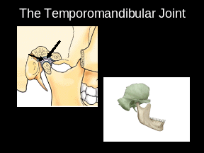
Notes:The mandible and cranium articulate at the temporomandibular joint (the TMJ). At rest, the head of the mandible is located in the glenoid fossa of the temporal bone. This is also called the mandibular fossa. The structure of the joint is unusual in that it contains a disc of fibrocartilage which separates the mandibular head from the temporal bone. Embryologically, this disc is derived from the same structure as the lateral pterygoid muscle, so the disc and the muscle are continuous with one another.
It’s important to understand that the movement of the mandibular head in the glenoid fossa as the mouth is opened and closed is not a simple rotation. Indeed, the head doesn’t even remain in the fossa as the mouth is opened. This is because it undergoes forward translation as well as rotation as the mouth opens. Take a look at this animation. You can see the mandibular head sliding forwards and backwards as the mouth opens and closes. Such sliding prevents interference between the mandible and more posterior structures as the mouth is opened, and it allows side-to-side movement of the mandible. If the mandibular head was limited to pure rotation, it would be impossible to swing the lower jaw from side-to-side, thereby reducing the ability of the teeth to grind up food.
|
|
Slide
5
The Lateral Pterygoid Muscle Duration: 00:01:40 Advance mode: By user |
|
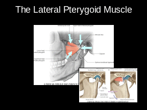
Notes:Let’s begin our tour of the muscles of mastication by looking at the lateral pterygoid muscle, which acts with the digastric muscle to open the mouth.
The lateral pterygoid muscle is a rather complicated structure as it has two heads which come together to insert into the neck of the mandible and parts of the TMJ.
The two heads of this muscle are called the upper head and the lower head. The anterior attachment of the upper head (which is, strictly speaking, called the origin of the upper head) is part of the sphenoid bone. The origin of the lower head is also part of the sphenoid bone, the lateral pterygoid plate. To be precise, the lower head takes its origin from the lateral surface of the lateral pterygoid plate. You should consult an anatomical atlas to see exactly where this is.
The two heads of the lateral pteryoid travel posteriorly, coming together just anterior to the neck of the mandible. The tendon of the muscle inserts into a depression on the neck of the mandible called the pterygoid fovea, and into the intra-articular disc and capsule of the TMJ.
As the fibres of the lateral pterygoid muscle run from an anterior origin (which is fixed) to a posterior insertion (which is mobile), when they contract, they pull the insertion site anteriorly. That is, they pull the neck of the mandible and parts of the TMJ (including the disc) forwards. This protrudes the mandible.
|
|
Slide
6
The Digastric Muscle Duration: 00:01:50 Advance mode: By user
Display : In Articulate player |
|
Notes:Of course, protruding the mandible isn’t what we think of when we ask someone to open their mouth. Another movement, rotation, is involved as well. The orientation of the lateral pterygoid muscle is such that it cannot be responsible for the rotatory movements that the mandible undergoes when the mouth is being opened. Somewhat ironically, the muscle that can be most closely associated with this rotation, the digastric, is not even classified as a muscle of mastication.
The digastric muscle takes its origin from the mastoid process, that large inferiorly projecting piece of the temporal bone you can feel just behind and below your external ear canal. The muscle is rather unusual in that it has two bellies – two fleshy pieces of muscular tissue – that are joined together by a tendon. This tendon runs through a loop of connective tissue joined to the hyoid bone in the upper part of the neck. Because of this unusual arrangement, contraction of the digastric’s muscular bellies draws the mandible downwards (that is, depresses it).
In this animation, you can see the movements that the mandibular head and intra-articular disc of the TMJ undergo as the mouth opens and closes. Unlike the very simple animation shown earlier, this 3-dimensional reconstruction shows the lateral pterygoid muscle pulling the mandibular head forwards during opening of the mouth. The muscle you can see just posterior to the mandible is the posterior belly of the digastric. This and the anterior belly contract in a co-ordinated manner with the lateral pterygoid to bring about depression and protrusion of the mandible. This is evident as opening of the mouth.
|
|
Slide
7
The Temporalis Muscle Duration: 00:00:58 Advance mode: By user |
|
Notes:The muscles involved in closing the mouth are the temporalis, the masseter and the medial pterygoid.
The temporalis is a large muscle originating on the lateral surface of the skull. It travels downwards deep to the zygomatic arch before inserting into the coronoid process of the mandible.
You can see that the most anterior fibres of the temporalis run almost vertically, whilst the posterior fibres have a much more horizontal orientation. This means that when temporalis contracts, it tends to elevate the mandible (mainly anterior fibres) whilst at the same time retracting it (mainly posterior fibres). Of course, this reverses the protrusion and depression that the mandible undergoes when the mouth is being opened.
|
|
Slide
8
The Masseter Muscle Duration: 00:00:41 Advance mode: By user
Display : In Articulate player |
|
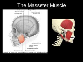
Notes:The temporalis acts in concert with two other muscles, the masseter and medial pterygoid when closing the mouth. Normally, the masseter is the most important of these two other muscles in this function.
The masseter muscle runs downwards from the zygomatic arch to insert into the outer surfaces of the mandibular ramus and coronoid process. Consequently, contraction of the masseter elevates the mandible, thereby assisting the temporalis in one of its two functions.
In this animation, you can see the temporalis and masseter working together when the mouth is being closed.
|
|
Slide
9
The Medial Pterygoid Muscle Duration: 00:02:04 Advance mode: By user
Display : In Articulate player |
|
Notes:The medial pterygoid runs from its origin (primarily the medial surface of the lateral pterygoid plate, though a smaller part of the muscle arises from the maxilla) to the inner aspect of the ramus of the mandible. If you look at the inner surface of the mandible, you can see a roughened area near the angle – this is the attachment point of the medial pterygoid.
The fibres of the medial pterygoid run infero-laterally from their origin to their insertion. This means that when they contract, they tend to pull the mandible upwards and medially. Contraction of just one medial pterygoid will tend to swing the mandible towards the opposite side of the face. Indeed, the main function of the medial pterygoid is in side-to-side movements of the mandible (assisted by the lateral pterygoid). You can see this in the animation. Of course, the initial movement in the animation would be brought about by the medial and lateral pterygoid muscles on the left-hand side of the head. Unfortunately, they are not shown in this animation so you only get to see the right-hand muscles returning the displaced mandible to the resting position.
You may be beginning to think that the actions of the muscles of mastication aren’t quite as simple as you originally thought. This is probably because you’re seeing that the muscles act together rather than as individuals. Thus, the lateral pterygoid can work with the digastric to open the mouth, and with the medial pterygoid to swing it from side to side. The lateral pterygoid contracts when you’re opening your mouth, but it also has a degree of contraction when you close it, in order to counter the effects of temporalis and masseter and thereby impart some finesse to the control of mouth closing.
|
|
Slide
10
Nerve Supplies Duration: 00:01:04 Advance mode: By user |
|
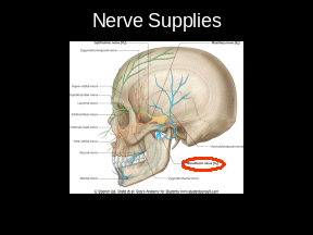
Notes:We’ve now met all of the muscles of mastication and their seemingly poor relative, the digastric. We’ve seen where they are and what they do. What we haven’t seen yet, it how they are controlled. For this, we need to know about the nerve supply to the muscles.
The good news is that all the muscles of mastication receive their motor and proprioceptive supply from the mandibular division of the trigeminal nerve (the fifth cranial nerve). This serves to re-enforce the importance of the trigeminal nerve for you as nerve V also provides the sensory supply to all the teeth and most of the gingivae.
The digastric muscle is rather different. Its anterior belly is supplied by a branch of the mandibular nerve like all the other muscles of mastication. In contrast, the posterior belly is supplied by a branch of the facial nerve, cranial nerve number VII.
|
|
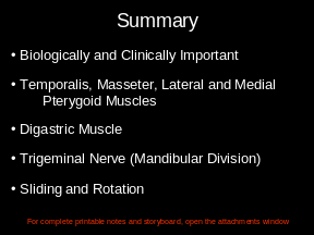
Slide
11
Summary Duration: 00:00:53 Advance mode: By user |
|
Notes:Today we’ve taken a look at the muscles of mastication.
We’ve seen that they play an important role in animal biology, and that they are of clinical importance for medical and dental practitioners.
We’ve seen that there are four muscles of mastication on each side of the head, the temporalis, masseter, lateral pterygoid and medial pterygoid.
The digastric muscle is important in opening the mouth, but is not classified as a muscle of mastication.
The motor and proprioceptive nerve inputs to the muscles of mastication are provided by the mandibular division of the trigeminal nerve.
The mandible does not undergo simple rotation during the opening and closing of the mouth. Rather, the mandibular head slides and rotates to produce the movements necessary for efficient mastication.
|
|
Slide 12 Quiz Duration: 00:00:05 Advance mode: By user |
|
Notes:
|
|
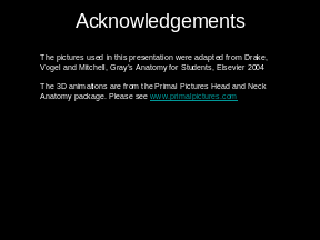
Slide 13 Acknowledgements Duration: 00:00:05 Advance mode: Auto |
|
Notes:The pictures used in this presentation were adapted from Drake, Vogel and Mitchell, Gray’s Anatomy for Students, Elsevier 2004
The 3D animations are from the Primal Pictures Head and Neck Anatomy package. Please see www.primalpictures.com
|


![]() Published
by Articulate™ Presenter www.articulate.com
Published
by Articulate™ Presenter www.articulate.com
Tags: details, 001330, slides, mastication, duration, presentation, presenter
- UNIVERSIDAD JOSÉ CARLOS MARIATEGUI MOQUEGUA AV EJERCITO MZ N
- BADANIE BENCHMARKINGOWE OBSZAR BADAWCZY 2 JAKOŚĆ NAPIĘCIA SUKCES BADAŃ
- Pravila in Režim Ribolovne Dovolilnice – Ujemi in Spusti
- BONES THEATRE 655 N OPPORTUNITY DRIVE COLUMBIA CITY IN
- HUMAN RESOURCES BRANCH ADVICE NO 0213 25 JUNE
- APPENDIX 1 KENNEDY VIETNAM AND THE PRESS THE “NEWS
- ZAŁĄCZNIK NR 2 DO REGULAMINU MAŁOPOLSKI KONKURS „MAM ZAWÓD
- CURRICULUM VITAE (CV) COMPLET INFORMATIONS REQUISES EXEMPLE IDENTIFICATION
- MINISTERIO DE VIVIENDA Y ASENTAMIENTOS HUMANOS CUESTIONARIO SOBRE
- PROTOCOLO DE EXPERIMENTO EFECTO DE ANESTÉSICOS LOCALES SOBRE LA
- FAULT TOLERANCE TODAY COMPUTERS ARE USED IN VERY COMPLEX
- RECURSOS HUMANOS PARA UNA COBERTURA SANITARIA UNIVERSAL UNA PLANTILLA
- Psihijatrijska Bolnica Ugljan Ulica Otočkih Dragovoljaca 42 23275 Ugljan
- ZWIĄZEK HARCERSTWA POLSKIEGO HUFIEC IM STANISŁAWA STASZICA W STARACHOWICACH
- UNA GENERACIÓN DE MARCAS PABLO GARCÍA RUIZ
- NEWS RELEASE WEEK OF FEBRUARY 19TH 2007 ANNE SCHELLMAN
- F ORM 511F MAY 9 2005 PAGE 4 OF
- THE GLOBAL CIVIL SOCIETY FORUM (GCSF) UNEPS GOVERNING COUNCIL
- la Coherencia y la Cohesión 1 la Coherencia
- T PÁGINA DEL TÍTULO IPO DE ARTÍCULO ORIGINAL TÍTULO
- STUDIEHANDBOK FÖR FORSKARSTUDERANDE VID AVDELNINGEN FÖR URBANA OCH REGIONALA
- SCOTT CUPIT & FI DAFF FROM MELBOURNE AUSTRALIA TRAVELLED
- CALENDARIO LABORAL 2021 EMPRESA DOMICILIO CENTRO DE TRABAJO CONVENIO
- CÓDIGO Nº COMISIÓN DE DESARROLLO DEL PREGRADO CODEPRE SOLICITUD
- WRITING STRONG PARAGRAPHS STRONG BODY PARAGRAPHS ARE THE KEY
- CNF SUPPLEMENT EFFECTIVE DATE SEPTEMBER 1 1996 DURATION THIS
- ÉSZREVÉTELEK A DOKTORI ISKOLÁKRÓL A DOKTORI ELJÁRÁSOK RENDJÉRŐL ÉS
- 1(1) BESTÄLLNING AV NYBYGGNADSKARTA FÖRENKLAD NYBYGGNADSKARTA UTDRAG UR PRIMÄRKARTA
- TABEL 51 TABEL 18 RENCANA PROGRAM KEGIATAN INDIKATOR
- 3H NUCLIDE SAFETY DATA SHEET HYDROGEN3 [TRITIUM] WWWNCHPSORG 3H
 DEBRECENI ORVOSTUDOMÁNYI EGYETEM OKTATÁSFEJLESZTÉSI KÖZPONT SEGÉDLET AZ ORVOSTANHALLGATÓK INFORMATIKAI
 LETRAS DEL DISCO “BSO DONKEY XOTE” A MI
VZOREC OBČINA GROSUPLJE TABORSKA CESTA 2 1290 GROSUPLJE KI
 ADJETIVOS DEMOSTRATIVOS VAN DELANTE DEL NOMBRE E INDICAN LA
21 SEMINARIO INTERNACIONAL DICTADURA Y SISTEMA REPRESIVO 19361948 UNIVERSIDAD
 PHRCPHREPHRL XXXX COURSE NAME SEMESTER YEAR FACULTY COORDINATOR(S) NAME
JAY ZARNIKAU JAY ZARNIKAU PHD RESEARCH FELLOW DEPARTMENT OF
 SPORTS CLUBSOCIETY OFFICER RECORD SHEET CLUBSOCIETY EFFECTIVE FROM (DATE)
2 LAMPIRAN XVIII PERATURAN KEPALA BADAN KOORDINASI PENANAMAN MODAL
УТВЪРЖДАВАМ П МАРТА АЛЕКСАНДРОВА ДРАКОВА ДИРЕКТОР ИД УПЪЛНОМОЩЕНО ЛИЦЕ
404! PAGE NOT FOUND THE REQUESTED URL SOMEURL WAS
HULL CITY COUNCIL ACCESSIBILITY STRATEGY FOR EDUCATIONAL SERVICES 2015
CUSTOMIZED LIVING (INCLUDING 24HOUR CUSTOMIZED LIVING) PAGE POSTED 10103
 ESPOL ICQA 1ERA EVALUACIÓN QUÍMICA GENERAL I
 STSGAC1040ADD2 NACIONES UNIDAS STSGAC1040ADD2 SECRETARÍA DISTR GENERAL 15 DE
ZAŁĄCZNIK NR 22 UMOWA KONTRAKTOWA O UDZIELENIE ZAMÓWIENIA NA
GIẤY XÁC NHẬN CHỈNH SỬA BÁO CÁO NGHIỆM THU
 P ARENT ARTICLE FOR NEWSLETTERS INEXPERIENCE AND WINTER ROADS
BP 4042 PERSONNEL GENERAL EMPLOYEE USE OF TECHNOLOGY THE
27 EXECUTIVE FUNCTIONS AND SCIENCE EXECUTIVE FUNCTIONS PREDICT CONCEPTUAL
